CROSS-REFERENCE TO RELATED APPLICATIONS
This application is the U.S. national stage pursuant to 35 U.S.C. §371, of United States international application Ser. No. PCT/US2012/064179, filed Nov. 8, 2012, designating the United States and published in English on May 16, 2013, a publication WO 2013/070933 A2, which claims the benefit of the following U.S. Provisional Application Ser. Nos. 61/556,850, filed Nov. 8, 2011, and 61/583,882, filed Jan. 6, 2012, the entire contents of which are incorporated herein by reference.
STATEMENT OF RIGHTS TO INVENTIONS MADE UNDER FEDERALLY SPONSORED RESEARCH
This work was supported by the following grant from the National Institutes of Health, Grant Nos.: CA89600 and CA86323. The government has certain rights in the invention.
BACKGROUND OF THE INVENTION
Prostate cancer is a leading healthcare concern in North America and Europe. There were an estimated 232,090 new cases of prostate cancer diagnosed in 2005 in the United States, and over 30,350 deaths from advanced metastatic disease. Prostate cancer is now the most commonly diagnosed lethal malignancy, and the second leading cause of cancer death of men in the United States. Although curative treatment (e.g., radical prostatectomy or radiotherapy) is feasible for many patients with the earliest stage disease, early diagnosis remains a challenge. If prostate cancer becomes metastatic, the median survival for such patients is approximately one year. There remains an urgent need for determining those at risk for or susceptible to prostate cancer, early-stage prostate cancer prognosis, and early intervention.
Prostate specific antigen (PSA) screening has led to earlier detection of PCA and significantly reduced PCA-associated fatalities. However, a major limitation of the serum PSA test is a lack of prostate cancer sensitivity and specificity especially in the intermediate range of PSA detection (4-10 ng/ml). Elevated serum PSA levels are often detected in patients with non-malignant conditions such as benign prostatic hyperplasia (BPH) and prostatitis, and provide little information about the aggressiveness of the cancer detected. Coincident with increased serum PSA testing, there has been a dramatic increase in the number of prostate needle biopsies performed. This has resulted in a surge of equivocal prostate needle biopsies. Thus, development of additional serum and tissue biomarkers or additional methods to detect a patient at risk for prostate cancer are urgently required.
SUMMARY OF THE INVENTION
As described below, the present invention features compositions and methods for the diagnosis, treatment and prevention of prostate cancer, as well as for treatment selection.
In one aspect, the invention provides a method for identifying a subject as having an increased propensity to develop prostate cancer, the method involving detecting an alteration in a HoxB13 nucleic acid sequence or amino acid sequence in a biological sample derived from the subject (e.g., where detecting an alteration by direct nucleic acid or amino acid sequencing, PCR, hybridization, TaqMan® probe, molecular beacon, FRET hybridization probe, 5′ nuclease probe, primer extension, Restriction Fragment Length Polymorphism (RFLP), mass spectrometry, or using a protein or nucleic acid microarray).
In another aspect, the invention provides a method of determining the prognosis of a subject identified as having prostate cancer, the method involving identifying an alteration in a HoxB13 nucleic acid sequence or amino acid sequence in a biological sample derived from the subject, where the subject is identified as having a positive family history for prostate cancer and is younger than age 55, thereby identifying the subject as having a poor prognosis relative to a reference subject.
In still another aspect, the invention provides a method for selecting a therapy for a subject identified as having prostate cancer, the method involving detecting an alteration in a HoxB13 nucleic acid sequence or amino acid sequence in a biological sample derived from the subject, where detection of such alteration is indicative that aggressive treatment (e.g., radical prostatectomy, radiation therapy, chemotherapy, hormone therapy, and/or androgen ablation) is required.
In another aspect, the invention provides an isolated HoxB13 nucleic acid molecule having one or more of the following mutations: a change of adenosine for guanine in the second position of codon 84 (GGA→GAA) resulting in a nonconservative substitution of glutamic acid for glycine (G84E); a missense mutation 685C→G resulting in the substitution of glycine for arginine at position 229 (R229G); a substitution of proline for leucine at codon 144 (431T→C); and a substitution of aspartic acid for tyrosine (Y88D) at codon 88 (262T→G).
In a related aspect, the invention provides an expression vector including a nucleic acid molecule according to any of the aspects described herein (e.g., having a promoter operably linked to a HoxB13 nucleic acid molecule). In another related aspect, the invention provides cell containing a vector according to any of the aspects described herein.
In another aspect, the invention provides an isolated nucleic acid molecule for detecting one or more of the following alterations in HOXB13: a change of adenosine for guanine in the second position of codon 84 (GGA→GAA) resulting in a nonconservative substitution of glutamic acid for glycine (G84E); a missense mutation 685C→G resulting in the substitution of glycine for arginine at position 229 (R229G); a substitution mutation 431T→C resulting in a substitution of leucine for proline (L144P); and a substitution of aspartic acid for tyrosine (Y88D) at codon 88 (262T→G).
In another aspect, the invention provides an isolated antibody that specifically binds a HoxB13 protein variant having Y88D, L144P, R229G, or G84E, but that does not specifically bind a wild-type HoxB13 protein.
In various embodiments of any of the aspects delineated herein, the alteration in HOXB13 is one or more of: a change of adenosine for guanine in the second position of codon 84 (GGA→GAA) resulting in a nonconservative substitution of glutamic acid for glycine (G84E); a missense mutation 685C→G resulting in the substitution of glycine for arginine at position 229 (R229G); a substitution of proline for leucine at codon 144 (431T→C); and a substitution of aspartic acid for tyrosine (Y88D) at codon 88 (262T→G).
In various embodiments of any of the aspects delineated herein, the subject is identified as having a positive family history for prostate cancer and is younger than age 55. In various embodiments of any of the aspects delineated herein, the G84E mutation is identified in a subject of Nordic descent. In various embodiments of any of the aspects delineated herein, the R229G mutation is identified in a subject of African-American descent. In various embodiments of any of the aspects delineated herein, the sample is a tissue sample, tissue biopsy sample, or biological liquid.
In various embodiments of any of the aspects delineated herein, the method identifies the subject as in need of increased surveillance for prostate disease (e.g., annual measurement of PSA levels in the subject. In various embodiments of any of the aspects delineated herein, the detection of increased PSA identifies the subject as having prostate cancer or in need of further testing.
In various embodiments of any of the aspects delineated herein, the nucleic acid molecule is suitable for amplification of the alteration. In various embodiments of any of the aspects delineated herein, the nucleic acid molecules hybridize or fail to hybridize to the mutant sequence. In various embodiments of any of the aspects delineated herein, the nucleic acid molecule includes a detectable moiety.
The invention provides compositions and methods for diagnosing, treating or preventing prostate cancer. Other features and advantages of the invention will be apparent from the detailed description, and from the claims.
DEFINITIONS
Unless defined otherwise, all technical and scientific terms used herein have the meaning commonly understood by a person skilled in the art to which this invention belongs. The following references provide one of skill with a general definition of many of the terms used in this invention: Singleton et al., Dictionary of Microbiology and Molecular Biology (2nd ed. 1994); The Cambridge Dictionary of Science and Technology (Walker ed., 1988); The Glossary of Genetics, 5th Ed., R. Rieger et al. (eds.), Springer Verlag (1991); and Hale & Marham, The Harper Collins Dictionary of Biology (1991). As used herein, the following terms have the meanings ascribed to them below, unless specified otherwise.
By “Homeobox B13 polypeptide” or “HOXB13 polypeptide” is meant a polypeptide or fragment thereof having at least 85% amino acid identity to NCBI Accession No. NP_006352 and having DNA binding activity.
By “HOXB13 nucleic acid molecule” is meant or a polynucleotide encoding a HOXB13 polypeptide. An exemplary HOXB13 nucleic acid molecule is provided at NCBI Accession No. NM_006361.
By “alteration” is meant any change in the nucleic acid or amino acid sequence of a molecule relative to a reference sequence. Such alteration may be a missense, frameshift or substitution mutation. The reference sequence is typically a wild-type HoxB13 nucleic acid or amino acid sequence.
As used herein, the term “antibody” means not only intact antibody molecules, but also fragments of antibody molecules that retain immunogen-binding ability.
By “biologic sample” is meant any tissue, cell, fluid, or other material derived from an organism.
By “clinical aggressiveness” is meant the severity of the neoplasia. Aggressive neoplasias are more likely to metastasize than less aggressive neoplasias. While conservative methods of treatment are appropriate for less aggressive neoplasias, more aggressive neoplasias require more aggressive therapeutic regimens.
By “detect” refers to identifying the presence, absence, level, or concentration of an agent.
By “detectable” is meant a moiety that when linked to a molecule of interest renders the latter detectable. Such detection may be via spectroscopic, photochemical, biochemical, immunochemical, or chemical means. For example, useful labels include radioactive isotopes, magnetic beads, metallic beads, colloidal particles, fluorescent dyes, electron-dense reagents, enzymes (for example, as commonly used in an ELISA), biotin, digoxigenin, or haptens.
By “genotype” is meant the genetic composition of a cell, organism, or individual. With reference to the invention, the genotype of an individual is determined as heterozygous or homozygous for one or more variant alleles of interest.
By “genotyping” is meant the characterization of the two alleles in one or more genes of interest (i.e., to determine a genotype).
By “heterozygous” is meant that a chromosomal locus has two different alleles. In one embodiment of the invention, heterozygous refers to a genotype in which one allele has a wild-type HOXB13 sequence and the other allele has a sequence encoding a HOXB13 variant that has an alteration at glycine 84 (e.g. G84E or rs138213197).
By “homozygous” is meant that a chromosomal locus has two identical alleles. In the invention, homozygous wild-type is meant to refer to a genotype in which both alleles have a wild-type HOXB13 sequence. In some embodiments, homozygous can refer to a genotype in which both alleles have a sequence encoding a HOXB13 variant that does not has an alteration at at glycine 84 (e.g. G84E or rs138213197).
By “increases” is meant a positive alteration of at least 10%, 25%, 50%, 75%, 100%, 200%, 300%, 400%, 500%, 1000%, or more.
By “propensity” is meant that a subject has an increased risk of developing disease relative to a reference subject. Such an increased risk is associated with the presence of an alteration in a HoxB13 nucleic acid or amino acid sequence that predisposes the subject to develop prostate cancer relative to the risk of prostate cancer in a reference subject carrying a wild-type HoxB13 sequence.
The terms “isolated,” “purified,” or “biologically pure” refer to material that is free to varying degrees from components which normally accompany it as found in its native state. “Isolate” denotes a degree of separation from original source or surroundings. “Purify” denotes a degree of separation that is higher than isolation. A “purified” or “biologically pure” protein is sufficiently free of other materials such that any impurities do not materially affect the biological properties of the protein or cause other adverse consequences. That is, a nucleic acid or peptide of this invention is purified if it is substantially free of cellular material, viral material, or culture medium when produced by recombinant DNA techniques, or chemical precursors or other chemicals when chemically synthesized. Purity and homogeneity are typically determined using analytical chemistry techniques, for example, polyacrylamide gel electrophoresis or high performance liquid chromatography. The term “purified” can denote that a nucleic acid or protein gives rise to essentially one band in an electrophoretic gel. For a protein that can be subjected to modifications, for example, phosphorylation or glycosylation, different modifications may give rise to different isolated proteins, which can be separately purified.
By “reference” is meant a standard of comparison. For example, the nucleotide sequence in a patient sample may be compared to the nucleotide sequence present in a corresponding healthy cell or tissue.
By “positive family history” is meant the presence of prostate cancer is a first degree relative (e.g., son, father, uncle, brother).
By “periodic” is meant at regular intervals. Periodic patient monitoring includes, for example, a schedule of tests that are administered daily, bi-weekly, bi-monthly, monthly, bi-annually, or annually.
By “severity of neoplasia” is meant the degree of pathology. The severity of a neoplasia increases, for example, as the stage or grade of the neoplasia increases.
By “marker” is meant any protein or polynucleotide having an alteration in activity, expression level, or sequence that is associated with a disease, disorder, or condition.
By “Marker profile” is meant a characterization of the expression or expression level of two or more polypeptides or polynucleotides.
As used herein a “nucleic acid or oligonucleotide probe” is defined as a nucleic acid capable of binding to a target nucleic acid of complementary sequence through one or more types of chemical bonds, usually through complementary base pairing, usually through hydrogen bond formation. As used herein, a probe may include natural (i.e., A, G, C, or T) or modified bases (7-deazaguanosine, inosine, etc.). In addition, the bases in a probe may be joined by a linkage other than a phosphodiester bond, so long as it does not interfere with hybridization. It will be understood by one of skill in the art that probes may bind target sequences lacking complete complementarity with the probe sequence depending upon the stringency of the hybridization conditions. The probes are preferably directly labeled with isotopes, for example, chromophores, lumiphores, chromogens, or indirectly labeled with biotin to which a streptavidin complex may later bind. By assaying for the presence or absence of the probe, one can detect the presence or absence of a target gene of interest.
By “reduces” is meant a negative alteration of at least 10%, 25%, 50%, 75%, or 100%.
By “reference” is meant a standard or control condition. In various embodiments of the invention, the reference is the wild-type sequence of a gene or gene isoform.
By “specifically binds” is meant a compound or antibody that recognizes and binds a polypeptide of the invention, but which does not substantially recognize and bind other molecules in a sample.
The phrase “selectively (or specifically) hybridizes to” refers to the binding, duplexing, or hybridizing of a molecule only to a particular nucleotide sequence under stringent hybridization conditions when that sequence is present in a complex mixture (for example, total cellular or library DNA or RNA).
By “single nucleotide polymorphism” or “SNP” is meant a DNA sequence variation occurring when a single nucleotide in the genome differs between members of a biological species or paired chromosomes in an individual. SNPs are used as genetic markers for variant alleles.
By “target nucleic acid molecule” is meant a nucleic acid or biomarker of the sample that is to be detected.
By “variant” as is meant a polynucleotide or polypeptide sequence that differs from a wild-type or reference sequence by one or more nucleotides or one or more amino acids. An exemplary HOXB13 variant includes HOXB13 (G84E or rs138213197).
Nucleic acid molecules useful in the methods of the invention include any nucleic acid molecule that encodes a polypeptide of the invention or a fragment thereof. Such nucleic acid molecules need not be 100% identical with an endogenous nucleic acid sequence, but will typically exhibit substantial identity. Polynucleotides having “substantial identity” to an endogenous sequence are typically capable of hybridizing with at least one strand of a double-stranded nucleic acid molecule. By “hybridize” is meant pair to form a double-stranded molecule between complementary polynucleotide sequences (e.g., a gene described herein), or portions thereof, under various conditions of stringency. (See, e.g., Wahl, G. M. and S. L. Berger (1987) Methods Enzymol. 152:399; Kimmel, A. R. (1987) Methods Enzymol. 152:507).
For example, stringent salt concentration will ordinarily be less than about 750 mM NaCl and 75 mM trisodium citrate, preferably less than about 500 mM NaCl and 50 mM trisodium citrate, and more preferably less than about 250 mM NaCl and 25 mM trisodium citrate. Low stringency hybridization can be obtained in the absence of organic solvent, e.g., formamide, while high stringency hybridization can be obtained in the presence of at least about 35% formamide, and more preferably at least about 50% formamide. Stringent temperature conditions will ordinarily include temperatures of at least about 30° C., more preferably of at least about 37° C., and most preferably of at least about 42° C. Varying additional parameters, such as hybridization time, the concentration of detergent, e.g., sodium dodecyl sulfate (SDS), and the inclusion or exclusion of carrier DNA, are well known to those skilled in the art. Various levels of stringency are accomplished by combining these various conditions as needed. In a preferred: embodiment, hybridization will occur at 30° C. C. in 750 mM NaCl, 75 mM trisodium citrate, and 1% SDS. In a more preferred embodiment, hybridization will occur at 37° C. C. in 500 mM NaCl, 50 mM trisodium citrate, 1% SDS, 35% formamide, and 100 .mu.g/ml denatured salmon sperm DNA (ssDNA). In a most preferred embodiment, hybridization will occur at 42° C. C. in 250 mM NaCl, 25 mM trisodium citrate, 1% SDS, 50% formamide, and 200 μg/ml ssDNA. Useful variations on these conditions will be readily apparent to those skilled in the art.
For most applications, washing steps that follow hybridization will also vary in stringency. Wash stringency conditions can be defined by salt concentration and by temperature. As above, wash stringency can be increased by decreasing salt concentration or by increasing temperature. For example, stringent salt concentration for the wash steps will preferably be less than about 30 mM NaCl and 3 mM trisodium citrate, and most preferably less than about 15 mM NaCl and 1.5 mM trisodium citrate. Stringent temperature conditions for the wash steps will ordinarily include a temperature of at least about 25° C., more preferably of at least about 42° C., and even more preferably of at least about 68° C. In a preferred embodiment, wash steps will occur at 25° C. in 30 mM NaCl, 3 mM trisodium citrate, and 0.1% SDS. In a more preferred embodiment, wash steps will occur at 42° C. in 15 mM NaCl, 1.5 mM trisodium citrate, and 0.1% SDS. In a more preferred embodiment, wash steps will occur at 68° C. in 15 mM NaCl, 1.5 mM trisodium citrate, and 0.1% SDS. Additional variations on these conditions will be readily apparent to those skilled in the art. Hybridization techniques are well known to those skilled in the art and are described, for example, in Benton and Davis (Science 196:180, 1977); Grunstein and Hogness (Proc. Natl. Acad. Sci., USA 72:3961, 1975); Ausubel et al. (Current Protocols in Molecular Biology, Wiley Interscience, New York, 2001); Berger and Kimmel (Guide to Molecular Cloning Techniques, 1987, Academic Press, New York); and Sambrook et al., Molecular Cloning: A Laboratory Manual, Cold Spring Harbor Laboratory Press, New York.
By “substantially identical” is meant a polypeptide or nucleic acid molecule exhibiting at least 50% identity to a reference amino acid sequence (for example, any one of the amino acid sequences described herein) or nucleic acid sequence (for example, any one of the nucleic acid sequences described herein). Preferably, such a sequence is at least 60%, more preferably 80% or 85%, and more preferably 90%, 95% or even 99% identical at the amino acid level or nucleic acid to the sequence used for comparison.
Sequence identity is typically measured using sequence analysis software (for example, Sequence Analysis Software Package of the Genetics Computer Group, University of Wisconsin Biotechnology Center, 1710 University Avenue, Madison, Wis. 53705, BLAST, BESTFIT, GAP, or PILEUP/PRETTYBOX programs). Such software matches identical or similar sequences by assigning degrees of homology to various substitutions, deletions, and/or other modifications. Conservative substitutions typically include substitutions within the following groups: glycine, alanine; valine, isoleucine, leucine; aspartic acid, glutamic acid, asparagine, glutamine; serine, threonine; lysine, arginine; and phenylalanine, tyrosine. In an exemplary approach to determining the degree of identity, a BLAST program may be used, with a probability score between e−3 and e−100 indicating a closely related sequence.
BRIEF DESCRIPTION OF THE DRAWINGS
FIG. 1 is a genetic pedigree chart showing the pedigrees of four subjects with the HOXB13 G84E Mutation on Initial Targeted Sequencing. The proband who was selected for sequencing is indicated by the arrow in each pedigree. The remaining symbols are described in the key. Squares indicate male sex, and circles female sex. Ages of subjects, rounded to the nearest 5-year interval, are shown under the symbols. A slash through the symbol indicates that the subject is deceased. Two subjects in two families, Family 1 from the University of Michigan Prostate Cancer Genetics Project (UM) and Family 1 from Johns Hopkins University (JHU), who were inferred to be obligate carriers of the HOXB13 G84E mutation, died from prostate cancer. The unaffected G84E carrier in JHU Family 1 was 70 years of age at last contact.
FIG. 2 depicts the structure of HOXB13. The HOXB13 gene is the most 5′ member of the HOXB gene cluster on chromosome 17q21-22. The locations of the five missense mutations are indicated in the two exons of HOXB13. The homeodomain region and MEIS interacting domains are indicated. CDS denotes coding sequences, and UTR untranslated regions.
FIG. 3 depicts DNA sequence chromatograms and associated histologic findings obtained from normal prostate and prostate-cancer tissue from a heterozygous carrier of the HOXB13 G84E variant. Wild-type and mutant DNA are present in both normal prostate tissue and prostate-cancer tissue from HOXB13 G84E carriers. DNA was extracted from sections of paraffin-embedded blocks of tissue obtained during a radical prostatectomy performed in a patient who was heterozygous for the HOXB13 G84E variant. The blocks were selected and trimmed to contain either normal or tumor tissue, as shown on hematoxylin and eosin staining (at left), and were subjected to Sanger sequencing. The chromatograms (at right) show the presence of both wild-type (GGA) and mutant (GAA) alleles at codon 84 in normal prostate tissue (middle) and ale maintenance of both alleles in the matched sample of prostate tumor tissue (bottom). The top chromatogram is a homozygous wild-type sequence (5′-GGTTACTTTGGAGGCGGG-3′ (SEQ ID NO: 4); translation: N-GYFGGG-C (SEQ ID NO: 5)) from a subject without the G84E mutation. The genome position shown (44,160,704) is based on the National Center for Biotechnology Information database, build 36 (hg18).
The New England Journal of Medicine
FIG. 4 depicts immunohistochemical staining for HOXB13 (Panels A, C, and E) and Alpha-methylacyl-CoA racemase (AMACR; Panels B and D) in benign (Panel A) and malignant prostate tissue (Panels B-E). Prominent staining of HOXB13 was observed in nuclei of both normal luminal epithelial cells and cancer cells. Tumor-specific staining of AMACR was present in the cytoplasm of cancer cells. Sections of Formalin-Fixed, Paraffin-Embedded (FFPE) tissue from a HOXB13 G84E carrier were stained with antibodies against HOXB13 (F-9, Santa Cruz Biotechnology) or AMACR (13H4, Dako North America Inc).
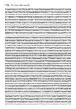
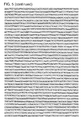
FIG. 5 provides genomic sequences of chromosome 17q21-22 (SEQ ID NO: 1). The HOXB13 G84E mutation is at chr 17 base pos. 46,805,705 GRCh37/hg19 (pos. 44,160,704 GRCh36/hg18). The DNA sequence surrounding this mutation is shown in FIG. 3. The GRCh37/hg19 coordinate for the Y88D mutation is at chr 17 base pos. 46,805,694; the L144P mutation is at 46,805,525; the G216C mutation is at 46,804,361; the R229G mutation is at 46,804,322.
DETAILED DESCRIPTION OF THE INVENTION
The invention features compositions and methods that are useful for the diagnosis, treatment and prevention of prostate cancer, as well as for treatment selection. The present invention is based, at least in part, on the discovery that having germline mutations in HOXB13 (e.g., G84E or rs138213197) increased the risk of developing prostate cancer. As reported in more detail below, the sequences of families having prostate cancer were analyzed at the 17q21-22 chromosomal locus to identify SNPs that were indicative of increased risk of prostate cancer. Despite a large degree of variability in the number of individuals sampled per pedigree, approximately 5% of prostate cancer families had at least one member with the G84E mutation (e.g., a germline mutation). Without being bound to a particular theory, the results are consistent with the hypothesis that HOXB13 G84E is a prostate cancer susceptibility allele that significantly increases the risk of prostate cancer. The identification and characterization of genetic variants reproducibly associated with substantial increases in prostate cancer risk would provide enhanced ability to identify men most likely to benefit from early disease screening.
Prostate Cancer
The development of human prostate cancer proceeds through a series of defined stages, beginning with prostatic intraepithelial neoplasia, progressing to invasive hormone-dependent cancer, and finally progressing to hormone-independent cancer. Most human prostate cancers are adenocarcinomas that express markers associated with luminal epithelial cells. Because of unbalanced cell proliferation, cell differentiation, and cell death, prostate cancer exhibits substantial histological heterogeneity. To date, DNA and tissue microarrays of tumors have failed to account for cellular heterogeneity and differences in the proliferative potential of different populations within tumors. At present, all of the phenotypically diverse cancer cells are treated as though they have unlimited proliferative potential and can acquire the ability to metastasize. In patients with metastic disease, conventional therapies are ineffective. Metastatic prostate tumor cells are able to survive extreme conditions within the circulation. Metastic cancer cells lodge in the capillary beds of distant organs where they undergo extensive proliferation, often in bone, lymph node, lung and brain [(Karhadkar et al., 2004; Swanson et al., 2006). Metastatic tumor cells share many characteristics (e.g., self-renewal, proliferation, and multi-potency) with pluripotent stem cells. Little is known about how human metastatic tumor cells maintain or acquire their multipotency. Recent studies suggest the existence of prostate cancer stem cells that are chemo-resistant and radiation-resistant. Therapies specifically directed against such cancer stem cells are likely to be more effective in curing prostate cancer and metastatic disease.
Accordingly, the present invention provides methods of treating prostate cancer and/or disorders or symptoms thereof which comprise administering a therapeutically effective amount of a pharmaceutical composition comprising an agent of the formulae herein to a subject (e.g., a mammal, such as a human). Thus, one embodiment is a method of treating a subject suffering from or susceptible to prostate cancer, metastatic prostate cancer, or prostate cancer having the propensity to metastasize or symptoms thereof. The method includes the step of administering to the mammal a therapeutic amount of an agent herein sufficient to treat the prostate cancer or symptom thereof, under conditions such that the prostate cancer is treated.
The methods herein include administering to the subject (including a subject identified as in need of such treatment) an effective amount of a compound described herein, or a composition described herein to produce such effect. Identifying a subject in need of such treatment can be in the judgment of a subject or a health care professional and can be subjective (e.g. opinion) or objective (e.g. measurable by a test or diagnostic method).
The therapeutic methods of the invention (which include prophylactic treatment) in general comprise administration of a therapeutically effective amount of the agents herein, such as an agent of the formulae herein to a subject (e.g., animal, human) in need thereof, including a mammal, particularly a human. Such treatment will be suitably administered to subjects, particularly humans, suffering from, having, susceptible to, or at risk for prostate cancer, including metastatic disease or prostate cancer having a propensity to metastasize, or a symptom thereof. Determination of those subjects “at risk” can be made by any objective or subjective determination by a diagnostic test or opinion of a subject or health care provider (e.g., genetic test, enzyme or protein marker, Marker (as defined herein), family history, and the like). The compounds herein may be also used in the treatment of any other disorders in which prostate cancer or hyperplasia may be implicated.
In one embodiment, the invention provides a method of monitoring treatment progress. The method includes the step of determining the presence of a SNP (e.g., or any target delineated herein modulated by a compound herein, a protein or indicator thereof, etc.) or diagnostic measurement (e.g., screen, assay) in a subject suffering from or susceptible to prostate cancer, in which the subject has been administered a therapeutic amount of a compound herein sufficient to treat the disease or symptoms thereof. The level of Marker determined in the method can be compared to known levels of Marker in either healthy normal controls or in other afflicted patients to establish the subject's disease status. In preferred embodiments, a second level of Marker in the subject is determined at a time point later than the determination of the first level, and the two levels are compared to monitor the course of disease or the efficacy of the therapy. In certain preferred embodiments, a pre-treatment level of Marker in the subject is determined prior to beginning treatment according to this invention; this pre-treatment level of Marker can then be compared to the level of Marker in the subject after the treatment commences, to determine the efficacy of the treatment.
Homeobox Protein B13 (Hox B13)
The HOX genes are a subfamily of the homeobox superfamily of transcription factors characterized by a highly conserved DNA-binding domain, or homeodomain. In humans, there are four HOX clusters, with each spanning approximately 200 kb on chromosomes 7 (HOXA), 17 (HOXB), 12 (HOXC), and 2 (HOXD). The combination of coordinated HOX expression provides a so-called HOX code that is essential for the pattern formation of the animal body. The genes within each HOX cluster are expressed temporally during development; 3′ genes are expressed early in anterior and proximal regions, whereas 5′ genes are expressed late in posterior and distal regions. HOX genes in paralogue group 13 are members of the abdominal B subfamily of such genes, which have posterior domains of expression, including in the developing urogenital system in vertebrates. Whereas multiple HOX13 paralogues are expressed during embryonic development of the prostate, HOXB13 maintains a high expression level into adulthood in normal prostate and, to a lesser level, in distal colon. In a study by Economides et al. (2003) mice that had been generated from embryonic stem cells with targeted disruption in HOXB13 had overgrowth of structures arising from the tail bud, including the spinal cord and tail vertebrae, with decreased apoptosis proposed as a possible mechanism. Further characterization of these animals showed subtle but definitive, lobe-specific abnormalities of the prostate gland but without evidence of preneoplastic lesions.
By sequencing coding regions of more than 200 genes in a previously identified region of linkage at 17q21-22, a rare but recurrent mutation (G84E) in HOXB13 was identified in four of 94 probands from hereditary prostate cancer families. The mutation co-segregated with prostate cancer in these four families and was found to be significantly more common among 5,083 unrelated prostate cancer patients (1.4%) than control subjects (0.1%) of Eu opean descent (p=8.5×10⁻⁷) leading to odds ratio (OR) estimates of 10-fold or more. In the studies described herein, the frequency of the mutation was higher in prostate cancer patients with early-onset disease (age at diagnosis=55 years old, 2.2%) or with a positive family history (2.2%), and most common in patients with both of these features (3.1%). Without being bound to a particular theory, these findings provide support for the concept that rare, moderately penetrant mutations as well as common, low-penetrance prostate cancer risk-associated variants identified from genome-wide association studies (GWAS) both contribute to prostate cancer risk.
The amino acid sequence of human HOXB13 is provided at NCBI Accession No. NP_006352, which is reproduced below:
| 1 |
mepgnyatld gakdiegllg agggrnlvah spltshpaap |
| |
| |
tlmpavnyap ldlpgsaepp |
| |
| 61 |
kqchpcpgvp qgtspapvpy gyfgggyysc rvsrsslkpc |
| |
| |
aqaatlaayp aetptageey |
| |
| 121 |
psrptefafy pgypgtyqpm asyldvsvvq tlgapgeprh |
| |
| |
dsllpvdsyq swalaggwns |
| |
| 181 |
qmccqgeqnp pgpfwkaafa dssgqhppda cafrrgrkkr |
| |
| |
ipyskgqlre lereyaankf |
| |
| 241 |
itkdkrrkis aatslserqi tiwfqnrrvk ekkvlakvkn |
| |
| |
satp |
The nucleotide sequence of an mRNA transcript encoding HOXB13 corresponds to NCBI Accession No. NM_130468 (human HOXB13 encoded at nucleotides 157-1011), which is reproduced below:
| 1 |
tcttgcgtca agacggccgt gctgagcgaa tgcaggcgac |
| |
| |
ttgcgagctg ggagcgattt |
| |
| 61 |
aaaacgcttt ggattccccc ggcctgggtg gggagagcga |
| |
| |
gctgggtgcc ccctagattc |
| |
| 121 |
cccgcccccg cacctcatga gccgaccctc ggctccatgg |
| |
| |
agcccggcaa ttatgccacc |
| |
| 181 |
ttggatggag ccaaggatat cgaaggcttg ctgggagcgg |
| |
| |
gaggggggcg gaatctggtc |
| |
| 241 |
gcccactccc ctctgaccag ccacccagcg gcgcctacgc |
| |
| |
tgatgcctgc tgtcaactat |
| |
| 301 |
gcccccttgg atctgccagg ctcggcggag ccgccaaagc |
| |
| |
aatgccaccc atgccctggg |
| |
| 361 |
gtgccccagg ggacgtcccc agctcccgtg ccttatggtt |
| |
| |
actttggagg cgggtactac |
| |
| 421 |
tcctgccgag tgtcccggag ctcgctgaaa ccctgtgccc |
| |
| |
aggcagccac cctggccgcg |
| |
| 481 |
taccccgcgg agactcccac ggccggggaa gagtacccca |
| |
| |
gccgccccac tgagtttgcc |
| |
| 541 |
ttctatccgg gatatccggg aacctaccag cctatggcca |
| |
| |
gttacctgga cgtgtctgtg |
| |
| 601 |
gtgcagactc tgggtgctcc tggagaaccg cgacatgact |
| |
| |
ccctgttgcc tgtggacagt |
| |
| 661 |
taccagtctt gggctctcgc tggtggctgg aacagccaga |
| |
| |
tgtgttgcca gggagaacag |
| |
| 721 |
aacccaccag gtcccttttg gaaggcagca tttgcagact |
| |
| |
ccagcgggca gcaccctcct |
| |
| 781 |
gacgcctgcg cctttcgtcg cggccgcaag aaacgcattc |
| |
| |
cgtacagcaa ggggcagttg |
| |
| 841 |
cgggagctgg agcgggagta tgcggctaac aagttcatca |
| |
| |
ccaaggacaa gaggcgcaag |
| |
| 901 |
atctcggcag ccaccagcct ctcggagcgc cagattacca |
| |
| |
tctggtttca gaaccgccgg |
| |
| 961 |
gtcaaagaga agaaggttct cgccaaggtg aagaacagcg |
| |
| |
ctacccctta agagatctcc |
| |
| 1021 |
ttgcctgggt gggaggagcg aaagtggggg tgtcctgggg |
| |
| |
agaccaggaa cctgccaagc |
| |
| 1081 |
ccaggctggg gccaaggact ctgctgagag gcccctagag |
| |
| |
acaacaccct tcccaggcca |
| |
| 1141 |
ctggctgctg gactgttcct caggagcggc ctgggtaccc |
| |
| |
agtatgtgca gggagacgga |
| |
| 1201 |
accccatgtg acagcccact ccaccagggt tcccaaagaa |
| |
| |
cctggcccag tcataatcat |
| |
| 1261 |
tcatcctgac agtggcaata atcacgataa ccagtactag |
| |
| |
ctgccatgat cgttagcctc |
| |
| 1321 |
atattttcta tctagagctc tgtagagcac tttagaaacc |
| |
| |
gctttcatga attgagctaa |
| |
| 1381 |
ttatgaataa atttggaagg cgatcccttt gcagggaagc |
| |
| |
tttctctcag acccccttcc |
| |
| 1441 |
attacacctc tcaccctggt aacagcagga agactgagga |
| |
| |
gaggggaacg ggcagattcg |
| |
| 1501 |
ttgtgtggct gtgatgtccg tttagcattt ttctcagctg |
| |
| |
acagctgggt aggtggacaa |
| |
| 1561 |
ttgtagaggc tgtctcttcc tccctccttg tccaccccat |
| |
| |
agggtgtacc cactggtctt |
| |
| 1621 |
ggaagcaccc atccttaata cgatgatttt tctgtcgtgt |
| |
| |
gaaaatgaag ccagcaggct |
| |
| 1681 |
gcccctagtc agtccttcct tccagagaaa aagagatttg |
| |
| |
agaaagtgcc tgggtaattc |
| |
| 1741 |
accattaatt tcctccccca aactctctga gtcttccctt |
| |
| |
aatatttctg gtggttctga |
| |
| 1801 |
ccaaagcagg tcatggtttg ttgagcattt gggatcccag |
| |
| |
tgaagtagat gtttgtagcc |
| |
| 1861 |
ttgcatactt agcccttccc aggcacaaac ggagtggcag |
| |
| |
agtggtgcca accctgtttt |
| |
| 1921 |
cccagtccac gtagacagat tcacagtgcg gaattctgga |
| |
| |
agctggagac agacgggctc |
| |
| 1981 |
tttgcagagc cgggactctg agagggacat gagggcctct |
| |
| |
gcctctgtgt tcattctctg |
| |
| 2041 |
atgtcctgta cctgggctca gtgcccggtg ggactcatct |
| |
| |
cctggccgcg cagcaaagcc |
| |
| 2101 |
agcgggttcg tgctggtcct tcctgcacct taggctgggg |
| |
| |
gtggggggcc tgccggcgca |
| |
| 2161 |
ttctccacga ttgagcgcac aggcctgaag tctggacaac |
| |
| |
ccgcagaacc gaagctccga |
| |
| 2221 |
gcagcgggtc ggtggcgagt agtggggtcg gtggcgagca |
| |
| |
gttggtggtg ggccgcggcc |
| |
| 2281 |
gccactacct cgaggacatt tccctcccgg agccagctct |
| |
| |
cctagaaacc ccgcggcggc |
| |
| 2341 |
cgccgcagcc aagtgtttat ggcccgcggt cgggtgggat |
| |
| |
cctagccctg tctcctctcc |
| |
| 2401 |
tgggaaggag tgagggtggg acgtgactta gacacctaca |
| |
| |
aatctattta ccaaagagga |
| |
| 2461 |
gcccgggact gagggaaaag gccaaagagt gtgagtgcat |
| |
| |
gcggactggg ggttcagggg |
| |
| 2521 |
aagaggacga ggaggaggaa gatgaggtcg atttcctgat |
| |
| |
ttaaaaaatc gtccaagccc |
| |
| 2581 |
cgtggtccag cttaaggtcc tcggttacat gcgccgctca |
| |
| |
gagcaggtca ctttctgcct |
| |
| 2641 |
tccacgtcct ccttcaagga agccccatgt gggtagcttt |
| |
| |
caatatcgca ggttcttact |
| |
| 2701 |
cctctgcctc tataagctca aacccaccaa cgatcgggca |
| |
| |
agtaaacccc ctccctcgcc |
| |
| 2761 |
gacttcggaa ctggcgagag ttcagcgcag atgggcctgt |
| |
| |
ggggaggggg caagatagat |
| |
| 2821 |
gagggggagc ggcatggtgc ggggtgaccc cttggagaga |
| |
| |
ggaaaaaggc cacaagaggg |
| |
| 2881 |
gctgccaccg ccactaacgg agatggccct ggtagagacc |
| |
| |
tttgggggtc tggaacctct |
| |
| 2941 |
ggactcccca tgctctaact cccacactct gctatcagaa |
| |
| |
acttaaactt gaggattttc |
| |
| 3001 |
tctgtttttc actcgcaata aattcagagc aaacaaaaaa |
| |
| |
aaaaaaa |
Several studies have examined the role of HOXB13 in normal and cancerous prostate biology, although substantially different conclusions have been reached, with HOXB13 being implicated as both a tumor suppressor and an oncogene in prostate and other cancers. For example, the growth of the prostate-cancer cell line LNCaP has been shown to be suppressed by both experimental overexpression of HOXB13 by transfection and by reduction of endogenous HOXB13 expression by RNA interference. HOXB13 physically interacts with the androgen receptor, one of the most important growth and differentiation regulators in normal and cancerous prostate biology. Without being bound to a particular theory, the HOXB13 G84E mutation is located in a conserved domain of the HOXB13 protein that has been shown to mediate binding to members of the MEIS protein family, which are implicated in leukemia. The studies described herein indicate that tumors in G84E carriers continue to express HOXB13 and maintain the mutant allele (FIGS. 3 and 4).
Diagnostic Assays
The present invention provides a number of diagnostic assays that are useful for the identification or characterization of prostate cancer in a subject. Such methods may be used alone or in combination with standard methods for monitoring a subject for prostate cancer. In one embodiment, a subject is identified as being at risk of developing prostate cancer by the presence of the SNP rs138213197 (corresponding to the genetic variant HOXB13 G84E), alone or in combination with other standard methods. To determine the stage or grade of a neoplasia, grading is used to describe how abnormal or aggressive the neoplastic cells appear, while staging is used to describe the extent of the neoplasia. If desired, the grade and stage of the neoplasia in combination with the presence of the SNP rs138213197 (corresponding to the genetic variant HOXB13 G84E) is used to determine a subject's long-term prognosis (i.e., probable response to treatment and survival). Thus, the methods of the invention are useful for predicting a patient's prognosis, and for selecting a course of treatment.
The Gleason scale is the most common scale used for grading prostate cancer. A pathologist will look at the two most poorly differentiated parts of the tumor and grade them. The Gleason score is the sum of the two grades, and so can range from two to 10. The higher the score is, the poorer the prognosis. Scores usually range between 4 and 7. The scores can be broken down into three general categories: (i) low-grade neoplasias (score<4) are typically slow-growing and contain cells that are most similar to normal prostate cells; intermediate grade neoplasias (4<score<7) are the most common and typically contain some cells that are similar to normal prostate cells as well as some more abnormal cells; high-grade neoplasias (8<score<10) contain cells that are most dissimilar to normal prostate cells. High-grade neoplasias are the most deadly because they are most aggressive and fast growing. High-grade neoplasias typically move rapidly into surrounding tissues, such as lymph nodes and bones.
Stage refers to the extent of a cancer. In prostate cancer, for example, one staging method divides the cancer into four categories, A, B, C, and D. Stage A describes a cancer that is only found by elevated PSA and biopsy, or at surgery for obstruction. It is not palpable on digital rectal exam (DRE). This stage is localized to the prostate. This type of cancer is usually curable, especially if it has a relatively low Gleason grade. Stage B refers to a cancer that can be felt on rectal examination and is limited to the prostate. Bone scans or CT/MRI scans are often used to determine this stage, particularly if prostate specific antigen (PSA) levels are significantly elevated or if the Gleason grade is 7 or greater. Many Stage B prostate cancers are curable. Stage C cancers have spread beyond the capsule of the prostate into local organs or tissues, but have not yet metastasized to other sites. This stage is determined by DRE, or CT/MRI scans, and/or sonography. In Stage C a bone scan or a PROSTASCINT scan is negative. Some Stage C cancers are curable. Stage D cancer has metastasized to distant lymph nodes, bones or other sites. This is usually determined by bone scan, PROSTASCINT scan, or other studies. Stage D cancer is usually incurable.
Types of Biological Samples
The presence of SNP rs138213197 (corresponding to the genetic variant HOXB13 G84E) can be detected in different types of biologic samples. In one embodiment, the biologic sample is a tissue sample that includes cells of a tissue or organ (e.g., prostatic tissue cells). Prostatic tissue is obtained, for example, from a biopsy of the prostate. In another embodiment, the biologic sample is a biologic fluid sample. Biological fluid samples include blood, blood serum, plasma, urine, seminal fluids, and ejaculate, or any other biological fluid useful in the methods of the invention.
Genotyping of HOXB13 Polymorphisms
A HOXB13 isoform is amplified by PCR to determine the genotype of the isoform, e.g., HOXB13 G84E. The amplified nucleic acid corresponding to HOXB13 may be analyzed using a variety of methods for detecting variant alleles to determine the genotype. The presence or absence of a polymorphism (e.g., G84E) in the HOXB13 gene may be evaluated using various techniques. For example, the HOXB13 gene is amplified by PCR and sequenced to determine the presence or absence of a single nucleotide polymorphism (SNP). In certain embodiments, real-time PCR may be used to detect a single nucleotide polymorphism of the amplified products. In other embodiments, a polymorphism in the amplified products may be detected using a technique including hybridization with a probe specific for a single nucleotide polymorphism, restriction endonuclease digestion, primer extension, microarray or gene chip analysis, mass spectrometry, or a DNAse protection assay.
Polymerase Chain Reaction (PCR)
Polymerase chain reaction (PCR) is widely known in the art. For example, U.S. Pat. Nos. 4,683,195, 4,683,202, and 4,800,159; K. Mullis, Cold Spring Harbor Symp. Quant. Biol., 51:263-273 (1986); and C. R. Newton & A. Graham, Introduction to Biotechniques: PCR, 2.sup.nd Ed., Springer-Verlag (New York: 1997), the disclosures of which are incorporated herein by reference, describe processes to amplify a nucleic acid sample target using PCR amplification extension primers which hybridize with the sample target. As the PCR amplification primers are extended, using a DNA polymerase (preferably thermostable), more sample target is made so that more primers can be used to repeat the process, thus amplifying the sample target sequence. Typically, the reaction conditions are cycled between those conducive to hybridization and nucleic acid polymerization, and those that result in the denaturation of duplex molecules.
In the first step of the reaction, the nucleic acid molecules of the sample are transiently heated, and then cooled, in order to denature double stranded molecules. Forward and reverse primers are present in the amplification reaction mixture at an excess concentration relative to the sample target. When the sample is incubated under conditions conducive to hybridization and polymerization, the primers hybridize to the complementary strand of the nucleic acid molecule at a position 3′ to the sequence of the region desired to be amplified that is the complement of the sequence whose amplification is desired. Upon hybridization, the 3′ ends of the primers are extended by the polymerase. The extension of the primer results in the synthesis of a DNA molecule having the exact sequence of the complement of the desired nucleic acid sample target. The PCR reaction is capable of exponentially amplifying the desired nucleic acid sequences, with a near doubling of the number of molecules having the desired sequence in each cycle. Thus, by permitting cycles of hybridization, polymerization, and denaturation, an exponential increase in the concentration of the desired nucleic acid molecule can be achieved.
The methods of the present invention involve amplifying regions of a polynucleotide with high fidelity using a thermostable DNA polymerase having 3′→5′ exonuclease activity. As defined herein, “3′→5′ exonuclease activity” refers to the activity of a template-specific nucleic acid polymerase having a 3′→5′ exonuclease activity associated with some DNA polymerases, in which one or more nucleotides are removed from the 3′ end of an oligonucleotide in a sequential manner. Polymerase enzymes having high fidelity 3′→5′ exonuclease activity are useful, for example, when primer extension must be performed over long distances (i.e., when the desired PCR amplification product is greater than about 5 kb). Polymerase enzymes having 3′→5′ exonuclease proofreading activity are known to those in the art. Examples of suitable proofreading enzymes include TaKaRa LA Taq (Takara Shuzo Co., Ltd.) and Pfu (Stratagene), Vent, Deep Vent (New England Biolabs). Exemplary methods for performing PCR are disclosed, for example, in U.S. Pat. No. 5,436,149; Barnes, Proc. Natl. Acad. Sci. USA 91:2216-2220 (1994); Tellier et al., Methods in Molecular Biology, Vol. 226, PCR Protocols, 2nd Edition, pp. 173-177; and, Cheng et al., Proc. Natl. Acad. Sci. 91:5695-5699 (1994); the contents of which are incorporated herein by reference. In various embodiments, PCR involves one DNA polymerase. In some embodiments, PCR may involve more than one DNA polymerase. When using a combination of polymerases in PCR, it is preferable to include one polymerase having 3′→5′ exonuclease activity, which assures high fidelity generation of the PCR product from the DNA template. Typically, a non-proofreading polymerase, which is the main polymerase is also used in conjunction with the proofreading polymerase in PCR reactions. PCR can also be performed using commercially available kits, such as LA PCR kit available from Takara Bio Inc.
Sequencing
DNA sequencing may be used to evaluate a polymorphism of the present invention. One DNA sequencing method is the Sanger method, which is also referred to as dideoxy sequencing or chain termination. The Sanger method is based on the use of dideoxynucleotides (ddNTP's) in addition to the normal nucleotides (NTP's) found in DNA. Dideoxynucleotides are essentially the same as nucleotides except they contain a hydrogen group on the 3′ carbon instead of a hydroxyl group (OH). These modified nucleotides, when integrated into a sequence, prevent the addition of further nucleotides. This occurs because a phosphodiester bond cannot form between the dideoxynucleotide and the next incoming nucleotide, and thus the DNA chain is terminated. Using this method, optionally coupled with amplification of the nucleic acid target, one can now rapidly sequence large numbers of target molecules, usually employing automated sequencing apparati. Such techniques are well known to those of skill in the art.
Pyrosequencing is another method of DNA sequencing that may be used to evaluate a polymorphism of the present invention, for example as described in U.S. Pat. Publ. No. 2006008824; herein incorporated by reference). Pyrosequencing, which is also referred to as sequencing by synthesis, involves taking a single strand of the DNA to be sequenced, synthesizing its complementary strand enzymatically one base pair at a time, and detecting by chemiluminescence the base that is added. In one embodiment, the template DNA is immobile, and solutions of A, C, G, and T nucleotides are sequentially added and removed from the reaction. Light is produced only when the nucleotide solution complements the first unpaired base of the template. The sequence of solutions which produce chemiluminescent signals allows the determination of the sequence of the template. The templates for pyrosequencing can be made both by solid phase template preparation (streptavidin-coated magnetic beads) and enzymatic template preparation (apyrase+exonuclease).
In a specific embodiment, ssDNA template is hybridized to a sequencing primer and incubated with the enzymes DNA polymerase, ATP sulfurylase, luciferase and apyrase, and with the substrates adenosine 5′ phosphosulfate (APS) and luciferin. The addition of one of the four deoxynucleotide triphosphates (dNTPs) (in place of dATP, dATPαS is added, which is not a substrate for a luciferase) initiates the second step. DNA polymerase incorporates the correct, complementary dNTPs onto the template, and the incorporation of the nucleotide releases pyrophosphate (PPi) stoichiometrically. ATP sulfurylase quantitatively converts PPi to ATP in the presence of adenosine 5′ phosphosulfate. The ATP generated acts to catalyze the luciferase-mediated conversion of luciferin to oxyluciferin and generates visible light in amounts that are proportional to the amount of ATP. The light produced in the luciferase-catalyzed reaction is detected by a camera and analyzed in a program. Unincorporated nucleotides and ATP are degraded by the apyrase, and the reaction can restart with another nucleotide.
Pyrosequencing, optionally coupled with amplification of the nucleic acid target, can sequence large numbers of target molecules, usually employing automated sequencing apparati, including long sequences (e.g., 400 million bp/10 hr in a single run).
Various PCR testing platforms that may be used with the present invention include: 5′ nuclease (TaqMan® probes), molecular beacons, and FRET hybridization probes. These detection methods rely on the transfer of light energy between two adjacent dye molecules, a process referred to as fluorescence resonance energy transfer (see, e.g., Espy et al (2006) Clin Microbiol Rev. 2006 January; 19(1): 165-256 for a review of various rtPCR approaches that may be used with the present invention).
5′ Nuclease Probes
In certain embodiments, a 5′ nuclease probe may be used to detect a polymorphism of the present invention. 5′ nuclease probes are often referred to by the proprietary name, TaqMan® probes. A TaqMan® probe is a short oligonucleotide (DNA) that contains a 5′ fluorescent dye and 3′ quenching dye. To generate a light signal (i.e., remove the effects of the quenching dye on the fluorescent dye), two events must occur. First, the probe must bind to a complementary strand of DNA, e.g., at about 60° C. Second, at this temperature, Taq polymerase, which is commonly used for PCR, must cleave the 5′ end of the TaqMan® probe (5′ nuclease activity), separating the fluorescent dye from the quenching dye.
In order to differentiate a single nucleotide polymorphism from a wild-type sequence in the DNA from a subject, a second probe with complementary nucleotide(s) to the polymorphism and a fluorescent dye with a different emission spectrum are typically utilized. Thus, these probes can be used to detect a specific, predefined polymorphism under the probe in the PCR amplification product. Two reaction vessels are typically used, one with a complementary probe to detect wild-type target DNA and another for detection of a specific nucleic acid sequence of a mutant strain. Because TaqMan® probes typically require temperatures of about 60° C. for efficient 5′ nuclease activity, the PCR may be cycled between about 90-95° C. and about 60° C. for amplification. In addition, the cleaved (free) fluorescent dye can accumulate after each PCR temperature cycle; thus, the dye can be measured at any time during the PCR cycling, including the hybridization step. In contrast, molecular beacons and FRET hybridization probes typically involve the measurement of fluorescence during the hybridization step.
Genotyping for the G84E polymorphism in the HOXB13 gene may be evaluated using the following (5′ endonuclease probe) real-time PCR technique. Genotyping assays can be performed in duplicate and analyzed on a Bio-Rad iCycler Iq® Multicolor Real-time detection system (Bio-Rad Laboratories, Hercules, Calif.). Real-time polymerase chain reaction (PCR) allelic discrimination assays to detect the presence or absence of specific single nucleotide polymorphisms in a HOXB13 gene, Gly143Glu (genomic: nt 9486; Cdna: nt 428) and Asp260fs (genomic: nt 12754; Cdna: nt 780), may utilize fluorogenic TaqMan® Probes.
Real-time PCR amplifications may be carried out in a 10 μl reaction mix containing 5 ng genomic DNA, 900 Nm of each primer, 200 Nm of each probe and 5 μl of 2× TaqMan® Universal PCR Master Mix (contains PCR buffer, passive reference dye ROX, deoxynucleotides, uridine, uracil-N-glycosylase and AmpliTaq Gold DNA polymerase; Perkin-Elmer, Applied Biosystems, Foster City, Calif.). Cycle parameters may be: 95° C. for 10 min, followed by 50 cycles of 92° C. for 15 sec and 60 C.° for 1 min. Real-time fluorescence detection can be performed during the 60° C. annealing/extension step of each cycle. The IQ software may be used to plot and automatically call genotypes based on a two parameter plot using fluorescence intensities of FAM and VIC at 49 cycles.
Molecular Beacons
Molecular beacons are another real-time PCR approach which may be used to identify the presence or absence of a polymorphism of the present invention. Molecular beacons are oligonucleotide probes that are labeled with a fluorescent dye (typically on the 5′ end) and a quencher dye (typically on the 3′ end). A region at each end of the molecular beacon probe is designed to be complementary to itself, so at low temperatures the ends anneal, creating a hairpin structure. This hairpin structure positions the two dyes in close proximity, quenching the fluorescence from the reporter dye. The central region of the probe is designed to be complementary to a region of a PCR amplification product. At higher temperatures, both the PCR amplification product and probe are single stranded. As the temperature of the PCR is lowered, the central region of the molecular beacon probe may bind to the PCR product and force the separation of the fluorescent reporter dye from the quenching dye. Without the quencher dye in close proximity, a light signal from the reporter dye can be detected. If no PCR amplification product is available for binding, the probe can re-anneal to itself, bringing the reporter dye and quencher dye into close proximity, thus preventing fluorescent signal.
Two or more molecular beacon probes with different reporter dyes may be used for detecting single nucleotide polymorphisms. For example, a first molecular beacon designed with a first reporter dye may be used to indicate the presence of a SNP and a second molecular beacon designed with a second reporter dye may be used to indicate the presence of the corresponding wild-type sequence; in this way, different signals from the first and/or second reporter dyes may be used to determine if a subject is heterozygous for a SNP, homozygous for a SNP, or homozygous wild-type at the corresponding DNA region. By selection of appropriate PCR temperatures and/or extension of the probe length, a molecular beacons may bind to a target PCR product when a nucleotide polymorphism is present but at a slight cost of reduced specificity. Molecular beacons advantageously do not require thermocycling, so temperature optimization of the PCR is simplified.
FRET Hybridization Probes
FRET hybridization probes, also referred to as LightCycler® probes, may also be used to detect a polymorphism of the present invention. FRET hybridization probes typically comprise two DNA probes designed to anneal next to each other in a head-to-tail configuration on the PCR product. Typically, the upstream probe has a fluorescent dye on the 3′ end and the downstream probe has an acceptor dye on the 5′ end. If both probes anneal to the target PCR product, fluorescence from the 3′ dye can be absorbed by the adjacent acceptor dye on the 5′ end of the second probe. As a result, the second dye is excited and can emit light at a third wavelength, which may be detected. If the two dyes do not come into close proximity in the absence of sufficient complimentary DNA, then FRET does not occur between the two dyes. The 3′ end of the second (downstream) probe may be phosphorylated to prevent it from being used as a primer by Taq during PCR amplification. The two probes may encompass a region of 40 to 50 DNA base pairs.
FRET hybridization probe technology permits melting curve analysis of the amplification product. If the temperature is slowly raised, probes annealing to the target PCR product will be reduced and the FRET signal will be lost. The temperature at which half the FRET signal is lost is referred to as the melting temperature of the probe system. A single nucleotide polymorphism in the target DNA under a hybridization FRET probe will still generate a signal, but the melting curve will display a lower Tm. The lowered Tm can indicate the presence of a specific polymorphism. The target PCR product is detected and the altered Tm informs the user there is a difference in the sequence being detected. Like molecular beacons, there is not a specific thermocycling temperature requirement for FRET hybridization probes. Like molecular beacons, FRET hybridization probes have the advantage of being recycled or conserved during PCR temperature cycling, and a fluorescent signal does not accumulate as PCR product accumulates after each PCR cycle.
Primer Extension
Primer extension is another technique which may be used according to the present invention. A primer and no more than three NTPs may be combined with a polymerase and the target sequence, which serves as a template for amplification. By using less than all four NTPs, it is possible to omit one or more of the polymorphic nucleotides needed for incorporation at the polymorphic site. It is important for the practice of the present invention that the amplification be designed such that the omitted nucleotide(s) is(are) not required between the 3′ end of the primer and the target polymorphism. The primer is then extended by a nucleic acid polymerase, in a preferred embodiment by Taq polymerase. If the omitted NTP is required at the polymorphic site, the primer is extended up to the polymorphic site, at which point the polymerization ceases. However, if the omitted NTP is not required at the polymorphic site, the primer will be extended beyond the polymorphic site, creating a longer product. Detection of the extension products is based on, for example, separation by size/length which will thereby reveal which polymorphism is present. For example, U.S. Ser. No. 10/407,846, which is which is hereby incorporated by reference, describes a form of primer extension.
RFLP
Restriction Fragment Length Polymorphism (RFLP) is a technique in which different DNA sequences may be differentiated by analysis of patterns derived from cleavage of that DNA. If two sequences differ in the distance between sites of cleavage of a particular restriction endonuclease, the length of the fragments produced will differ when the DNA is digested with a restriction enzyme. The similarity of the patterns generated can be used to differentiate species (and even strains) from one another.
Restriction endonucleases in turn are the enzymes that cleave DNA molecules at specific nucleotide sequences depending on the particular enzyme used. Enzyme recognition sites are usually 4 to 6 base pairs in length. Generally, the shorter the recognition sequence, the greater the number of fragments generated. If molecules differ in nucleotide sequence, fragments of different sizes may be generated. The fragments can be separated by gel electrophoresis. Restriction enzymes are isolated from a wide variety of bacterial genera and are thought to be part of the cell's defenses against invading bacterial viruses. Use of RFLP and restriction endonucleases in SNP analysis requires that the SNP affect cleavage of at least one restriction enzyme site.
Mass Spectrometry
Mass spectrometry may also be used to detect a polymorphism of the present invention. By exploiting the intrinsic properties of mass and charge, mass spectrometry (MS) can resolved and confidently identified a wide variety of complex compounds. Traditional quantitative MS has used electrospray ionization (ESI) followed by tandem MS (MS/MS) (Chen et al., 2001; Thong et al., 2001; Wu et al., 2000) while newer quantitative methods are being developed using matrix assisted laser desorption/ionization (MALDI) followed by time of flight (TOF) MS (Bucknall et al., 2002; Mirgorodskaya et al., 2000; Gobom et al., 2000). Methods of mass spectroscopy that may be used with the present invention include: ESI, ESI tandem mass spectroscopy (ESI/MS/MS), Secondary ion mass spectroscopy (SIMS), Laser desorption mass spectroscopy (LD-MS), Laser Desorption Laser Photoionization Mass Spectroscopy (LDLPMS), and MALDI-TOF-MS.
Hybridization
There are a variety of ways by which one can assess genetic profiles, and may of these rely on nucleic acid hybridization. Hybridization is defined as the ability of a nucleic acid to selectively form duplex molecules with complementary stretches of DNAs and/or RNAs. Depending on the application envisioned, one would employ varying conditions of hybridization to achieve varying degrees of selectivity of the probe or primers for the target sequence.
Typically, a probe or primer of between 13 and 100 nucleotides, preferably between 17 and 100 nucleotides in length up to 1-2 kilobases or more in length will allow the formation of a duplex molecule that is both stable and selective. Such fragments may be readily prepared, for example, by directly synthesizing the fragment by chemical means or by introducing selected sequences into recombinant vectors for recombinant production.
For applications requiring high selectivity, one will typically desire to employ relatively high stringency conditions to form the hybrids. For example, relatively low salt and/or high temperature conditions, such as provided by about 0.02 M to about 0.10 M NaCl at temperatures of about 50° C. to about 70° C. Such high stringency conditions tolerate little, if any, mismatch between the probe or primers and the template or target strand and would be particularly suitable for isolating specific genes or for detecting specific mRNA transcripts. It is generally appreciated that conditions can be rendered more stringent by the addition of increasing amounts of formamide.
For certain applications, for example, lower stringency conditions may be used. Under these conditions, hybridization may occur even though the sequences of the hybridizing strands are not perfectly complementary, but are mismatched at one or more positions. Conditions may be rendered less stringent by increasing salt concentration and/or decreasing temperature. For example, a medium stringency condition could be provided by about 0.1 to 0.25 M NaCl at temperatures of about 37° C. to about 55° C., while a low stringency condition could be provided by about 0.15 M to about 0.9 M salt, at temperatures ranging from about 20° C. to about 55° C. Hybridization conditions can be readily manipulated depending on the desired results.
In other embodiments, hybridization may be achieved under conditions of, for example, 50 mM Tris-HCl (pH 8.3), 75 mM KCl, 3 mM MgCl2, 1.0 mM dithiothreitol, at temperatures between approximately 20° C. to about 37° C. Other hybridization conditions utilized could include approximately 10 mM Tris-HCl (pH 8.3), 50 mM KCl, 1.5 mM MgCl2, at temperatures ranging from approximately 40° C. to about 72° C.
In certain embodiments, it will be advantageous to employ nucleic acids of defined sequences of the present invention in combination with an appropriate means, such as a label, for determining hybridization. A wide variety of appropriate indicator means are known in the art, including fluorescent, radioactive, enzymatic or other ligands, such as avidin/biotin, which are capable of being detected. In preferred embodiments, one may desire to employ a fluorescent label or an enzyme tag such as urease, alkaline phosphatase or peroxidase, instead of radioactive or other environmentally undesirable reagents. In the case of enzyme tags, colorimetric indicator substrates are known that can be employed to provide a detection means that is visibly or spectrophotometrically detectable, to identify specific hybridization with complementary nucleic acid containing samples.
In general, it is envisioned that the probes or primers described herein will be useful as reagents in solution hybridization, as in PCR, for detection of expression of corresponding genes, as well as in embodiments employing a solid phase. In embodiments involving a solid phase, the test DNA (or RNA) is adsorbed or otherwise affixed to a selected matrix or surface. This fixed, single-stranded nucleic acid is then subjected to hybridization with selected probes under desired conditions. The conditions selected will depend on the particular circumstances (depending, for example, on the G+C content, type of target nucleic acid, source of nucleic acid, size of hybridization probe, etc.). Optimization of hybridization conditions for the particular application of interest is well known to those of skill in the art. After washing of the hybridized molecules to remove non-specifically bound probe molecules, hybridization is detected, and/or quantified, by determining the amount of bound label. Representative solid phase hybridization methods are disclosed in U.S. Pat. Nos. 5,843,663, 5,900,481 and 5,919,626. Other methods of hybridization that may be used in the practice of the present invention are disclosed in U.S. Pat. Nos. 5,849,481, 5,849,486 and 5,851,772. The relevant portions of these and other references identified in this section of the Specification are incorporated herein by reference.
Microarrays
The invention provides diagnostic microarrays for detecting the SNP rs138213197 (corresponding to the genetic variant HOXB13 G84E) in a biological sample. HOXB13 nucleic acid molecules or polypeptides are useful as hybridizable array elements in the microarray. The array elements are organized in an ordered fashion such that each element is present at a specified location on the substrate. Useful substrate materials include membranes, composed of paper, nylon or other materials, filters, chips, glass slides, and other solid supports. The ordered arrangement of the array elements allows hybridization patterns and intensities to be interpreted as expression levels of particular genes or proteins. Methods for making nucleic acid microarrays are known to the skilled artisan and are described, for example, in U.S. Pat. No. 5,837,832, Lockhart, et al. (Nat. Biotech. 14:1675-1680, 1996), and Schena, et al. (Proc. Natl. Acad. Sci. 93:10614-10619, 1996), herein incorporated by reference. Methods for making polypeptide microarrays are described, for example, by Ge (Nucleic Acids Res. 28:e3.i-e3.vii, 2000), MacBeath et al., (Science 289:1760-1763, 2000), Zhu et al. (Nature Genet. 26:283-289), and in U.S. Pat. No. 6,436,665, hereby incorporated by reference.
Nucleic Acid Microarrays
To produce a nucleic acid microarray oligonucleotides may be synthesized or bound to the surface of a substrate using a chemical coupling procedure and an ink jet application apparatus, as described in PCT application WO95/251116 (Baldeschweiler et al.), incorporated herein by reference. Alternatively, a gridded array may be used to arrange and link cDNA fragments or oligonucleotides to the surface of a substrate using a vacuum system, thermal, UV, mechanical or chemical bonding procedure.
A nucleic acid molecule (e.g. RNA or DNA) derived from a biological sample may be used to produce a hybridization probe as described herein. The biological samples are generally derived from a patient, preferably as a bodily fluid (such as blood, cerebrospinal fluid, phlegm, saliva, or urine) or tissue sample (e.g. a tissue sample obtained by biopsy). For some applications, cultured cells (e.g., lymphocytes) or other tissue preparations may be used. The mRNA is isolated according to standard methods, and cDNA is produced and used as a template to make complementary RNA suitable for hybridization. Such methods are described herein. The RNA is amplified in the presence of fluorescent nucleotides, and the labeled probes are then incubated with the microarray to allow the probe sequence to hybridize to complementary oligonucleotides bound to the microarray.
Incubation conditions are adjusted such that hybridization occurs with precise complementary matches or with various degrees of less complementarity depending on the degree of stringency employed. For example, stringent salt concentration will ordinarily be less than about 750 mM NaCl and 75 mM trisodium citrate, preferably less than about 500 mM NaCl and 50 mM trisodium citrate, and most preferably less than about 250 mM NaCl and 25 mM trisodium citrate. Low stringency hybridization can be obtained in the absence of organic solvent, e.g., formamide, while high stringency hybridization can be obtained in the presence of at least about 35% formamide, and most preferably at least about 50% formamide. Stringent temperature conditions will ordinarily include temperatures of at least about 30° C., more preferably of at least about 37° C., and most preferably of at least about 42° C. Varying additional parameters, such as hybridization time, the concentration of detergent, e.g., sodium dodecyl sulfate (SDS), and the inclusion or exclusion of carrier DNA, are well known to those skilled in the art. Various levels of stringency are accomplished by combining these various conditions as needed. In a preferred embodiment, hybridization will occur at 30° C. in 750 mM NaCl, 75 mM trisodium citrate, and 1% SDS. In a more preferred embodiment, hybridization will occur at 37° C. in 500 mM NaCl, 50 mM trisodium citrate, 1% SDS, 35% formamide, and 100 .mu.g/ml denatured salmon sperm DNA (ssDNA). In a most preferred embodiment, hybridization will occur at 42° C. in 250 mM NaCl, 25 mM trisodium citrate, 1% SDS, 50% formamide, and 200 .mu.g/ml ssDNA. Useful variations on these conditions will be readily apparent to those skilled in the art.
The removal of nonhybridized probes may be accomplished, for example, by washing. The washing steps that follow hybridization can also vary in stringency. Wash stringency conditions can be defined by salt concentration and by temperature. As above, wash stringency can be increased by decreasing salt concentration or by increasing temperature. For example, stringent salt concentration for the wash steps will preferably be less than about 30 mM NaCl and 3 mM trisodium citrate, and most preferably less than about 15 mM NaCl and 1.5 mM trisodium citrate. Stringent temperature conditions for the wash steps will ordinarily include a temperature of at least about 25° C., more preferably of at least about 42° C., and most preferably of at least about 68° C. In a preferred embodiment, wash steps will occur at 25° C. in 30 mM NaCl, 3 mM trisodium citrate, and 0.1% SDS. In a more preferred embodiment, wash steps will occur at 42° C. in 15 mM NaCl, 1.5 mM trisodium citrate, and 0.1% SDS. In a most preferred embodiment, wash steps will occur at 68° C. in 15 mM NaCl, 1.5 mM trisodium citrate, and 0.1% SDS. Additional variations on these conditions will be readily apparent to those skilled in the art.
A detection system may be used to measure the absence, presence, and amount of hybridization for all of the distinct sequences simultaneously (e.g., Heller et al., Proc. Natl. Acad. Sci. 94:2150-2155, 1997). Preferably, a scanner is used to determine the levels and patterns of fluorescence.
Protein Microarrays
Proteins, such as those described herein, may also be analyzed using protein microarrays. Such arrays are useful in high-throughput low-cost screens to identify peptide or candidate compounds that bind a polypeptide of the invention, or fragment thereof. Typically, protein microarrays feature a protein, or fragment thereof, bound to a solid support. Suitable solid supports include membranes (e.g., membranes composed of nitrocellulose, paper, or other material), polymer-based films (e.g., polystyrene), beads, or glass slides. For some applications, proteins (e.g., polypeptides encoded by a nucleic acid molecule listed at table 2 or Table 4 or antibodies against such polypeptides) are spotted on a substrate using any convenient method known to the skilled artisan (e.g., by hand or by inkjet printer). Preferably, such methods retain the biological activity or function of the protein bound to the substrate (Ge et al., supra; Zhu et al., supra).
The protein microarray is hybridized with a detectable probe. Such probes can be polypeptide, nucleic acid, or small molecules. For some applications, polypeptide and nucleic acid probes are derived from a biological sample taken from a patient, such as a bodily fluid (such as blood, urine, saliva, or phlegm); a homogenized tissue sample (e.g. a tissue sample obtained by biopsy); or cultured cells (e.g., prostate cancer cells). Probes can also include antibodies, candidate peptides, nucleic acids, or small molecule compounds derived from a peptide, nucleic acid, or chemical library. Hybridization conditions (e.g., temperature, pH, protein concentration, and ionic strength) are optimized to promote specific interactions. Such conditions are known to the skilled artisan and are described, for example, in Harlow, E. and Lane, D., Using Antibodies: A Laboratory Manual. 1998, New York: Cold Spring Harbor Laboratories. After removal of non-specific probes, specifically bound probes are detected, for example, by fluorescence, enzyme activity (e.g., an enzyme-linked calorimetric assay), direct immunoassay, radiometric assay, or any other suitable detectable method known to the skilled artisan.
Antibodies
Antibodies that selectively bind a variant HOXB13 polypeptide (e.g., G84E) are useful in the methods of the invention. Methods of preparing antibodies are well known to those of ordinary skill in the science of immunology. As used herein, the term “antibody” means not only intact antibody molecules, but also fragments of antibody molecules that retain immunogen-binding ability. Such fragments are also well known in the art and are regularly employed both in vitro and in vivo. Accordingly, as used herein, the term “antibody” means not only intact immunoglobulin molecules but also the well-known active fragments F(ab′)2, and Fab. F(ab′)2, and Fab fragments that lack the Fc fragment of intact antibody, clear more rapidly from the circulation, and may have less non-specific tissue binding of an intact antibody (Wahl et al., J. Nucl. Med. 24:316-325 (1983). The antibodies of the invention comprise whole native antibodies, bispecific antibodies; chimeric antibodies; Fab, Fab′, single chain V region fragments (scFv), fusion polypeptides, and unconventional antibodies.
In one embodiment, the antibody is a polyclonal antibody. The preparation and use of polyclonal antibodies are known to the skilled artisan. The invention also encompasses hybrid antibodies, in which one pair of heavy and light chains is obtained from a first antibody, while the other pair of heavy and light chains is obtained from a different second antibody. Such hybrids may also be formed using humanized heavy and light chains. Such antibodies are often referred to as “chimeric” antibodies.
In general, intact antibodies are said to contain “Fc” and “Fab” regions. The Fc regions are involved in complement activation and are not involved in antigen binding. An antibody from which the Fc′ region has been enzymatically cleaved, or which has been produced without the Fc′ region, designated an “F(ab′)2” fragment, retains both of the antigen binding sites of the intact antibody. Similarly, an antibody from which the Fc region has been enzymatically cleaved, or which has been produced without the Fc region, designated an “Fab′” fragment, retains one of the antigen binding sites of the intact antibody. Fab′ fragments consist of a covalently bound antibody light chain and a portion of the antibody heavy chain, denoted “Fd.” The Fd fragments are the major determinants of antibody specificity (a single Fd fragment may be associated with up to ten different light chains without altering antibody specificity). Isolated Fd fragments retain the ability to specifically bind to immunogenic epitopes.
Antibodies can be made by any of the methods known in the art utilizing a HOXB13 polypeptide (e.g., G84E), or fragments thereof. One method of obtaining antibodies is to immunize suitable host animals with an immunogen and to follow standard procedures for polyclonal or monoclonal antibody production. The immunogen will facilitate presentation of the immunogen on the cell surface. Immunization of a suitable host can be carried out in a number of ways. Nucleic acid sequences encoding a polypeptide involved in chondroitin sulfate biosynthesis or immunogenic fragments thereof, can be provided to the host in a delivery vehicle that is taken up by immune cells of the host. The cells will in turn express the receptor on the cell surface generating an immunogenic response in the host. Alternatively, nucleic acid sequences encoding a HOXB13 polypeptide (e.g., G84E), or immunogenic fragments thereof, can be expressed in cells in vitro, followed by isolation of the polypeptide and administration of the polypeptide to a suitable host in which antibodies are raised.
Alternatively, antibodies may, if desired, be derived from an antibody phage display library. A bacteriophage is capable of infecting and reproducing within bacteria, which can be engineered, when combined with human antibody genes, to display human antibody proteins. Phage display is the process by which the phage is made to ‘display’ the human antibody proteins on its surface. Genes from the human antibody gene libraries are inserted into a population of phage. Each phage carries the genes for a different antibody and thus displays a different antibody on its surface. Antibodies made by any method known in the art can then be purified from the host. Antibody purification methods may include salt precipitation (for example, with ammonium sulfate), ion exchange chromatography (for example, on a cationic or anionic exchange column preferably run at neutral pH and eluted with step gradients of increasing ionic strength), gel filtration chromatography (including gel filtration HPLC), and chromatography on affinity resins such as protein A, protein G, hydroxyapatite, and anti-immunoglobulin.
Antibodies can be conveniently produced from hybridoma cells engineered to express the antibody. Methods of making hybridomas are well known in the art. The hybridoma cells can be cultured in a suitable medium, and spent medium can be used as an antibody source. Polynucleotides encoding the antibody of interest can in turn be obtained from the hybridoma that produces the antibody, and then the antibody may be produced synthetically or recombinantly from these DNA sequences. For the production of large amounts of antibody, it is generally more convenient to obtain an ascites fluid. The method of raising ascites generally comprises injecting hybridoma cells into an immunologically naive histocompatible or immunotolerant mammal, especially a mouse. The mammal may be primed for ascites production by prior administration of a suitable composition (e.g., Pristane).
Monoclonal antibodies (Mabs) produced by methods of the invention can be “humanized” by methods known in the art. “Humanized” antibodies are antibodies in which at least part of the sequence has been altered from its initial form to render it more like human immunoglobulins. Techniques to humanize antibodies are particularly useful when non-human animal (e.g., murine) antibodies are generated. Examples of methods for humanizing a murine antibody are provided in U.S. Pat. Nos. 4,816,567, 5,530,101, 5,225,539, 5,585,089, 5,693,762 and 5,859,205.
In other embodiments, the invention provides “unconventional antibodies.” Unconventional antibodies include, but are not limited to, nanobodies, linear antibodies (Zapata et al., Protein Eng. 8(10): 1057-1062, 1995), single domain antibodies, single chain antibodies, and antibodies having multiple valencies (e.g., diabodies, tribodies, tetrabodies, and pentabodies). Nanobodies are the smallest fragments of naturally occurring heavy-chain antibodies that have evolved to be fully functional in the absence of a light chain. Nanobodies have the affinity and specificity of conventional antibodies although they are only half of the size of a single chain Fv fragment. The consequence of this unique structure, combined with their extreme stability and a high degree of homology with human antibody frameworks, is that nanobodies can bind therapeutic targets not accessible to conventional antibodies. Recombinant antibody fragments with multiple valencies provide high binding avidity and unique targeting specificity to cancer cells. These multimeric scFvs (e.g., diabodies, tetrabodies) offer an improvement over the parent antibody since small molecules of ˜60-100 kDa in size provide faster blood clearance and rapid tissue uptake See Power et al., (Generation of recombinant multimeric antibody fragments for tumor diagnosis and therapy. Methods Mol Biol, 207, 335-50, 2003); and Wu et al. (Anti-carcinoembryonic antigen (CEA) diabody for rapid tumor targeting and imaging. Tumor Targeting, 4, 47-58, 1999).
Various techniques for making unconventional antibodies have been described. Bispecific antibodies produced using leucine zippers are described by Kostelny et al. (J. Immunol. 148(5):1547-1553, 1992). Diabody technology is described by Hollinger et al. (Proc. Natl. Acad. Sci. USA 90:6444-6448, 1993). Another strategy for making bispecific antibody fragments by the use of single-chain Fv (sFv) diners is described by Gruber et al. (J. Immunol. 152:5368, 1994). Trispecific antibodies are described by Tutt et al. (J. Immunol. 147:60, 1991). Single chain Fv polypeptide antibodies include a covalently linked VH::VL heterodimer which can be expressed from a nucleic acid including VH- and VL-encoding sequences either joined directly or joined by a peptide-encoding linker as described by Huston, et al. (Proc. Nat. Acad. Sci. USA, 85:5879-5883, 1988). See, also, U.S. Pat. Nos. 5,091,513, 5,132,405 and 4,956,778; and U.S. Patent Publication Nos. 20050196754 and 20050196754.
Patient Monitoring
After a subject is identified as at risk of developing prostate cancer (e.g., by the detection of SNP rs138213197 (corresponding to the genetic variant HOXB13 G84E), the subject may be monitored for the development of prostate cancer. Typically, those at risk of developing prostate cancer have a familial history of prostate cancer. In men of European or Nordic descent, the detection of SNP rs138213197 (corresponding to the genetic variant HOXB13 G84E) is particularly informative, and this test is initially performed at an early age (e.g. at about 20, 25, 30, 35, 40, 45 yrs of age or younger). Subjects testing positive for the SNP rs138213197 under the age of 55, should be actively monitored for the development of prostate cancer once a year or more (e.g. annually). Tests for monitoring an individual identified at risk of developing prostate cancer includes measurement of levels of a marker (e.g., PSA) in the subject or a biological sample obtained from the subject. Such monitoring may be useful, for example, in assessing the onset of cancer in a patient. In prostate cancer, curative treatment (e.g., radical prostatectomy or radiotherapy) by early stage detection correlates with positive outcomes.
Selection of a Treatment Method
After a subject is diagnosed as having prostate cancer, a method of treatment is selected. In prostate cancer, for example, a number of standard treatment regimens are available. The detection of SNP rs138213197 (corresponding to the genetic variant HOXB13 G84E) in a biological sample may be used in selecting a treatment method. In one embodiment, less aggressive neoplasias may not have the SNP rs138213197 (corresponding to the genetic variant HOXB13 G84E) compared to more aggressive neoplasias. In one embodiment, a neoplastic cell that has a SNP rs138213197 (corresponding to the genetic variant HOXB13 G84E) correlates with a poor clinical outcome, such as metastasis or death. In other embodiments, the presence of SNP rs138213197 (corresponding to the genetic variant HOXB13 G84E) is indicative of a poor prognosis, i.e., identifying the prostate cancer as an aggressive prostate cancer. The absence of SNP rs138213197 (corresponding to the genetic variant HOXB13 G84E) correlates with a good clinical outcome. Such prostate cancers are identified as less aggressive.
While methods of neoplasia treatment vary depending on the type of neoplasia, the stage of neoplasia, and the patient's age, health, and physical condition, more aggressive treatment regimens will be used in patients having a poor prognosis (e.g., patients having a metastatic prostate carcinoma or a prostate carcinoma with metastatic potential). As described above, the methods of the invention are useful in determining the prognosis of a patient having neoplasia, such as a neoplasia with increased metastatic potential. In such patients aggressive therapies may be used. These include therapies having increased toxicity and those having an increased risk of adverse side-effects. Aggressive therapies are employed earlier and at higher doses in patients having a poor prognosis.
Less aggressive prostate cancers are likely to be susceptible to conservative treatment methods. Conservative treatment methods include, for example, cancer surveillance, which involves periodic patient monitoring using diagnostic assays of the invention, alone or in combination, with PSA blood tests and DREs, or hormonal therapy. Cancer surveillance is selected when diagnostic assays indicate that the adverse effects of treatment (e.g., impotence, urinary, and bowel disorders) are likely to outweigh therapeutic benefits.
More aggressive neoplasias are less susceptible to conservative treatment methods. When methods of the invention indicate that a neoplasia is very aggressive, an aggressive method of treatment should be selected. Aggressive therapeutic regimens typically include one or more of the following therapies: radical prostatectomy, radiation therapy (e.g., external beam and brachytherapy), hormone therapy, and chemotherapy.
Methods of the invention may be used alone or in combination with conservative or aggressive therapeutic regimens to treat a prostate cancer. While methods of neoplasia treatment vary depending on the type of neoplasia, the stage of neoplasia, and the patient's age, health, and physical condition, more aggressive treatment regimens will be used in patients having a poor prognosis (e.g., patients having a metastatic prostate carcinoma or a prostate carcinoma with metastatic potential). As described above, the methods of the invention are useful in determining the prognosis of a patient having neoplasia, such as a neoplasia with increased metastatic potential. In such patients aggressive therapies may be used. These include therapies having increased toxicity and those having an increased risk of adverse side-effects. Aggressive therapies are employed earlier and at higher doses in patients having a poor prognosis.
Standard neoplasia therapies, which methods are known to the skilled artisan (e.g., Wadler et al., Cancer Res. 50:3473-86, 1990), include, but are not limited to, chemotherapy, hormone therapy, androgen ablation, immunotherapy (include, but are not limited to, immunotherapy that will specifically target cancer stem cell transcription factors), radiotherapy, cryotherapy, surgery (e.g., radical prostatectomy), and any other therapeutic method used for the treatment of neoplasia. Prostate cancer depends in part on androgenic signaling for growth and survival. Androgens exert their cellular and physiologic effects through binding to the androgen receptor (AR), a member of the steroid hormone receptor family of transcription factors. Androgen binding to the AR ligand binding domain allows entry of the ligand-bound receptor into the nucleus and subsequent transcriptional regulation of androgen-responsive genes. Androgen ablation suppresses or blocks the production or action of male sex hormones for the treatment of cancers that rely upon male hormones for growth (e.g., removal of the testicles removed, taking female sex hormones, or taking antiandrogens). Hormonal therapy has been used since 1941 for the treatment of metastatic prostate cancer. Hormone deprivation therapies employing surgical and/or medical castration as well as their combination with anti-androgens have since become the mainstay of systemic treatment for advanced prostate cancer. Hormonal therapies for advanced prostate cancer target AR-mediated functions by suppressing the production of androgens and/or androgen binding to the AR ligand binding domain.
Treatment may include administration of one or more chemotherapeutics typically used in the treatment of a neoplasm, such as abiraterone acetate, altretamine, anhydrovinblastine, auristatin, bexarotene, bicalutamide, BMS184476, 2,3,4,5,6-pentafluoro-N-(3-fluoro-4-methoxyphenyl)benzene sulfonamide, bleomycin, N,N-dimethyl-L-valyl-L-valyl-N-methyl-L-valyl-L-proly-1-Lproline-t-butylamide, cachectin, cemadotin, chlorambucil, cyclophosphamide, 3′,4′-didehydro-4′-deoxy-8′-norvin-caleukoblastine, docetaxol, doxetaxel, cyclophosphamide, carboplatin, carmustine (BCNU), cisplatin, cryptophycin, cyclophosphamide, cytarabine, dacarbazine (DTIC), dactinomycin, daunorubicin, dolastatin, doxorubicin (adriamycin), etoposide, 5-fluorouracil, finasteride, flutamide, hydroxyurea and hydroxyureataxanes, ifosfamide, liarozole, lonidamine, lomustine (CCNU), mechlorethamine (nitrogen mustard), melphalan, mivobulin isethionate, rhizoxin, sertenef, streptozocin, mitomycin, methotrexate, 5-fluorouracil, nilutamide, onapristone, paclitaxel, prednimustine, procarbazine, RPR109881, stramustine phosphate, tamoxifen, tasonermin, taxol, tretinoin, vinblastine, vincristine, vindesine sulfate, and vinflunine. Other examples of chemotherapeutic agents can be found in Cancer Principles and Practice of Oncology by V. T. Devita and S. Hellman (editors), 6th edition (Feb. 15, 2001), Lippincott Williams & Wilkins Publishers.
Compounds of the present invention may be administered by any appropriate route for the treatment or prevention of neoplasia. These may be administered to humans, domestic pets, livestock, or other animals with a pharmaceutically acceptable diluent, carrier, or excipient, in unit dosage form. Administration may be parenteral, intravenous, intra-arterial, subcutaneous, intramuscular, intracranial, intraorbital, ophthalmic, intraventricular, intracapsular, intraspinal, intracisternal, intraperitoneal, intranasal, aerosol, by suppositories, or oral administration.
Therapeutic formulations may be in the form of liquid solutions or suspensions; for oral administration, formulations may be in the form of tablets or capsules; and for intranasal formulations, in the form of powders, nasal drops, or aerosols. Methods well known in the art for making formulations are found, for example, in Remington: The Science and Practice of Pharmacy (20th ed., ed. A. R. Gennaro, 2000, Lippincott Williams & Wilkins). Formulations for parenteral administration may, for example, contain excipients, sterile water, or saline, polyalkylene glycols such as polyethylene glycol, oils of vegetable origin, or hydrogenated napthalenes. Biocompatible, biodegradable lactide polymer, lactide/glycolide copolymer, or polyoxyethylene-polyoxypropylene copolymers may be used to control the release of the compounds. Nanoparticulate formulations (e.g., biodegradable nanoparticles, solid lipid nanoparticles, liposomes) may be used to control the biodistribution of the compounds. Other potentially useful parenteral delivery systems include ethylene-vinyl acetate copolymer particles, osmotic pumps, implantable infusion systems, and liposomes. Formulations for inhalation may contain excipients, for example, lactose, or may be aqueous solutions containing, for example, polyoxyethylene-9-lauryl ether, glycholate and deoxycholate, or may be oily solutions for administration in the form of nasal drops, or as a gel. The concentration of the compound in the formulation will vary depending upon a number of factors, including the dosage of the drug to be administered, and the route of administration.
The compound may be optionally administered as a pharmaceutically acceptable salt, such as a non-toxic acid addition salts or metal complexes that are commonly used in the pharmaceutical industry. Examples of acid addition salts include organic acids such as acetic, lactic, pamoic, maleic, citric, malic, ascorbic, succinic, benzoic, palmitic, suberic, salicylic, tartaric, methanesulfonic, toluenesulfonic, or trifluoroacetic acids or the like; polymeric acids such as tannic acid, carboxymethyl cellulose, or the like; and inorganic acid such as hydrochloric acid, hydrobromic acid, sulfuric acid phosphoric acid, or the like. Metal complexes include zinc, iron, and the like.
Administration of compounds in controlled release formulations is useful where the compound of formula I has (i) a narrow therapeutic index (e.g., the difference between the plasma concentration leading to harmful side effects or toxic reactions and the plasma concentration leading to a therapeutic effect is small; generally, the therapeutic index, TI, is defined as the ratio of median lethal dose (LD50) to median effective dose (ED50)); (ii) a narrow absorption window in the gastro-intestinal tract; or (iii) a short biological half-life, so that frequent dosing during a day is required in order to sustain the plasma level at a therapeutic level.
Many strategies can be pursued to obtain controlled release in which the rate of release outweighs the rate of metabolism of the therapeutic compound. For example, controlled release can be obtained by the appropriate selection of formulation parameters and ingredients, including, e.g., appropriate controlled release compositions and coatings. Examples include single or multiple unit tablet or capsule compositions, oil solutions, suspensions, emulsions, microcapsules, microspheres, nanoparticles, patches, and liposomes.
Formulations for oral use include tablets containing the active ingredient(s) in a mixture with non-toxic pharmaceutically acceptable excipients. These excipients may be, for example, inert diluents or fillers (e.g., sucrose and sorbitol), lubricating agents, glidants, and antiadhesives (e.g., magnesium stearate, zinc stearate, stearic acid, silicas, hydrogenated vegetable oils, or talc). Formulations for oral use may also be provided as chewable tablets, or as hard gelatin capsules wherein the active ingredient is mixed with an inert solid diluent, or as soft gelatin capsules wherein the active ingredient is mixed with water or an oil medium.
Kits
The invention also provides kits for genotyping a HOXB13 gene, particularly for the treatment or prevention of prostate cancer having a HOXB13 germline mutation (e.g., SNP rs138213197 corresponding to the genetic variant HOXB13 G84E). Such kits are useful for the diagnosis of a sequence alteration in HOXB13 relative to wild-type HOXB13 in a biological sample obtained from a subject. Alternatively, the invention provides for selecting a drug treatment regimen or adjusting a dosage. In various embodiments, the kit includes at least one primer pair that identifies a HOXB13 nucleic acid sequence, together with instructions for using the primers to genotype in a biological sample. In additional embodiments, the kit also includes instructions for selecting an appropriate therapy for a subject, monitoring drug therapy in a subject, identifying a subject as responsive to drug therapy, or identifying a subject as sensitive to a drug. Advantageously, such testing is carried out prior to drug administration or after an adverse event associated with drug administration. Preferably, the primers are provided in combination with a thermostable DNA polymerase capable of long-range PCR amplification (e.g., a high density array). In yet another embodiment, the kit further comprises a pair of primers capable of binding to and amplifying a reference sequence. In yet other embodiments, the kit comprises a sterile container which contains the primers; such containers can be boxes, ampules, bottles, vials, tubes, bags, pouches, blister-packs, or other suitable container form known in the art. Such containers can be made of plastic, glass, laminated paper, metal foil, or other materials suitable for holding nucleic acids.
The practice of the present invention employs, unless otherwise indicated, conventional techniques of molecular biology (including recombinant techniques), microbiology, cell biology, biochemistry and immunology, which are well within the purview of the skilled artisan. Such techniques are explained fully in the literature, such as, “Molecular Cloning: A Laboratory Manual”, second edition (Sambrook, 1989); “Oligonucleotide Synthesis” (Gait, 1984); “Animal Cell Culture” (Freshney, 1987); “Methods in Enzymology” “Handbook of Experimental Immunology” (Weir, 1996); “Gene Transfer Vectors for Mammalian Cells” (Miller and Calos, 1987); “Current Protocols in Molecular Biology” (Ausubel, 1987); “PCR: The Polymerase Chain Reaction”, (Mullis, 1994); “Current Protocols in Immunology” (Coligan, 1991). These techniques are applicable to the production of the polynucleotides and polypeptides of the invention, and, as such, may be considered in making and practicing the invention. Particularly useful techniques for particular embodiments will be discussed in the sections that follow.
The following examples are put forth so as to provide those of ordinary skill in the art with a complete disclosure and description of how to make and use the assay, screening, and therapeutic methods of the invention, and are not intended to limit the scope of what the inventors regard as their invention.
EXAMPLES
Example 1
Targeted Sequencing of 202 Candidate Genes Identified an SNP in HOXB13, a Change of Adenosine for Guanine (Transition, c.251G→A), Associated with Risk of Developing Prostate Cancer
Sequence data was reviewed for the presence of nonsense or missense mutations in 202 genes in the genetic region of interest (chromosome 17q21-22) (Lange et al., 2007). Exons (2009 exons) were sequenced from genes in the chromosome 17 candidate interval. The average depth of coverage across all loci was 49.5X. There were a total of 20 loci that yielded an average depth of coverage <1X (<1%), while 2040 loci had an average depth of coverage >10X (97%). On average 705 variants per sample were detected across the target region. Approximately 694 variants on average were present in dbSNP134 (98%), with an average of ˜12 novel variants per individual. The sequencing results are shown at Table 1.
Probands from four families were observed to have the same nonsynonymous mutation in HOXB13, a change of adenosine for guanine (transition, c.251G→A) in the second position of codon 84 (GGA→GAA), resulting in a nonconservative substitution of glutamic acid for glycine (G84E). At the time of this analysis, this mutation, now identified as rs138213197, was not reported in dbSNP, the database of known DNA sequence variants of the National Center for Biotechnology Information (NCBI), nor in the May 2011 release of the NCBI 1000 Genomes sequencing project, which included 1094 subjects, including 381 of European descent.
Given the importance and specificity of HOXB13 in prostate biology, further characterization of this mutation was performed. DNA samples from family members from each of these four pedigrees were sequenced to identify carriers of the HOXB13 G84E mutation. Cosegregation of the mutation with disease was observed in all 18 affected men with available DNA in the four families (FIG. 1).
Example 2
A HOXB13 G84E Mutation is Associated with Risk of Developing Prostate Cancer
To further assess HOXB13 G84E as a prostate cancer susceptibility allele, several additional populations of European descent were studied with a total of 5083 unrelated case subjects and 1401 unrelated control subjects. Clinical characteristics of genotyped samples are shown at Table 2.
The case subjects included a cohort of 1130 patients with early-onset or familial prostate cancer from the Prostate Cancer Genetics Project, 161 patients with hereditary prostate cancer from Johns Hopkins University, a series of 3499 men with localized prostate cancer who were treated with radical prostatectomy at Johns Hopkins Hospital, and 293 men who were treated for advanced prostate cancer at Johns Hopkins Hospital; the control population of 1401 men were found to have no prostate cancer during screening. Among men in the latter screening group, the G84E
| TABLE 1 |
| |
| Complete HOXB13 Sequencing Results from 94 Men with Prostate Cancer |
| Chrm 17 |
Ref Allele/Variant |
|
|
|
# Individuals |
| Location* |
Allele |
dbSNP ID |
Variant Type |
Substitution |
[N(%)] |
| |
| HOXB13 variants identified in 85 Caucasian samples |
| 44157313 |
G/T |
rs116931900**
|
3′ UTR |
NA |
7 (8.2) |
| 44157328 |
T/C |
rs79812861**
|
3′ UTR |
NA |
5 (5.9) |
| 44157843 |
G/C |
novel |
3′ UTR |
NA |
1 (1.2) |
| 44158537 |
C/T |
rs11653611 |
3′ UTR |
NA |
51 (60.0) |
| 44160442 |
A/G |
rs9900627 |
Synonymous |
S171S |
13 (15.3) |
| 44160589 |
G/A |
rs8556 |
Synonymous |
S122S |
20 (23.5) |
| 44160704 |
C/T |
rs138213197** |
Nonsynonymous |
G84E |
4 (4.7) |
| 44161086 |
T/C |
novel |
5′ UTR |
NA |
2 (2.4) |
| HOXB13 variants identified in 7 African American samples |
| 44157328 |
T/C |
rs79812861**
|
3′ UTR |
NA |
1 (14.3) |
| 44158537 |
C/T |
rs11653611 |
3′ UTR |
NA |
2 (28.6) |
| 44159094 |
C/T |
rs141179592** |
3′ UTR |
NA |
1 (14.3) |
| 44159321 |
G/C |
novel |
Nonsynonymous |
R229G |
1 (14.3) |
| 44160442 |
A/G |
rs9900627 |
Synonymous |
S171S |
2 (28.6) |
| 44160589 |
G/A |
rs8556 |
Synonymous |
S122S |
5 (71.4) |
| HOXB13 variants identified in 2 Asian American samples |
| 44157328 |
T/C |
rs79812861** |
3′ UTR |
NA |
2 (100) |
| 44158537 |
C/T |
rs11653611 |
3′ UTR |
NA |
2 (100) |
| 44160589 |
G/A |
rs8556 |
Synonymous |
S122S |
2 (100) |
| |
| *Location based on hg18 reference sequence |
| **These dbSNP ID numbers were not available at the completion of sequencing. |
| TABLE 2 |
| |
| Clinical features of genotyped samples. |
| All men studied described themselves as European descent and the sample |
| sizes are variable due to missing data. |
| | Mean | St. Dev. | Median | Range |
| |
| Age at Dx (yrs) (n = 1130) | 52.2 | 6.3 | 52 | 27-77 |
| RP Gleason Grade (n = 938) | 6.60 | 0.85 | 7 | 3-10 |
| Age at Dx (yrs) (n = 3797) | 58.5 | 6.7 | 59 | 35-85 |
| RP Gleason Grade (n = 3752) | 6.47 | 0.79 | 6 | 4-10 |
| |
| Abbreviations: RP = radical prostatectomy, Dx = diagnosis, St. Dev. = standard deviation |
mutation was found in only 1 man, resulting in a carrier-frequency estimate of 0.1%. Homozygous carriers were not identified among either case or control subjects.
Overall, men with prostate cancer were significantly more likely to carry the HOXB13 G84E allele (carrier frequency, 1.4%) than were those without prostate cancer (carrier frequency, 0.1%) (P=8.5×10⁻⁷; odds ratio, 20.1; 95% confidence interval [CI], 3.5 to 803.3). HOXB13 G84E mutation in sets of prostate cancer cases are shown at Table 3.
The carrier frequency varied as a function of age at diagnosis and family history, with the highest rates among men with both a positive family history and early diagnosis (≦55 years of age). The carrier frequency in this group (3.1%) was significantly higher than in men with early-onset prostate cancer who did not have a family history of the disease (1.0%, P=0.002) or in men with a family history in whom prostate cancer was diagnosed after the age of 55 years (1.2%, P=0.004). Frequencies of HOXB13 G84E carriers in prostate cancer cases of European descent stratified by family history and age at diagnosis are shown at Table 4.
The lowest carrier frequency was observed in men in whom prostate cancer was diagnosed after the age of 55 years and who did not have a family history, although this frequency was still higher than in control subjects (0.6%; odds ratio, 8.7; 95% CI, 1.2 to 381.3; P=0.02). A summary of results comparing age at diagnosis and family history strata to controls are shown at Table 5.
| TABLE 3 |
| |
| Summary of HOXB13 G84E mutation in sets of prostate cancer cases and controls of European descent |
| |
G84E |
Non- |
Carrier |
|
|
|
|
| |
Carriers |
Carriers |
Frequency |
p-value# |
Odds Ratio# |
p-value## |
Odds Ratio## |
| |
| 85 UM-PCGP and JHU |
4 |
81 |
4.9 |
4.8 × 10⁻⁵ |
68.4 |
5.4 × 10⁻⁵ |
32.6 |
| Sequenced HPC Families |
|
|
|
|
|
|
|
| All UM-PCGP and JHU |
72 |
5011 |
1.4 |
8.5 × 10⁻⁷ |
20.1 |
2.4 × 10⁻⁹ |
9.5 |
| Cases* |
|
|
|
|
|
|
|
| UM PCGP Data |
|
|
|
|
|
|
|
| UM-PCGP Early-onset and |
26 |
1104 |
2.3 |
1.0 × 10⁻⁸ |
33.0 |
1.3 × 10⁻¹⁰ |
15.6 |
| HPC* Cases |
|
|
|
|
|
|
|
| Johns Hopkins Data |
|
|
|
|
|
|
|
| JHU HPC*, Prostatectomy |
46 |
3907 |
1.2 |
1.6 × 10⁻⁵ |
16.5 |
6.0 × 10⁻⁷ |
7.8 |
| and Oncology Cases |
|
|
|
|
|
|
|
| Controls | |
|
|
|
|
|
|
| JHU Controls |
| |
1 |
1400 |
0.071 |
n.a. |
n.a. |
n.a. |
n.a. |
| Exome Sequencing Project + |
3 |
1258 |
0.24 |
n.a. |
n.a. |
n.a. |
n.a. |
| CEU HapMap |
| |
| *Unrelated cases (the case with earliest age at diagnosis selected from HPC families) excluding 85 European-descent families used for discovery. |
| #Comparison to 1401 JHU controls. |
| ##Comparison to 1401 JHU controls + 1235 Exome Sequencing Project subjects + 28 CEU unrelated genotyped subjects. |
| TABLE 4 |
| |
| Frequencies of HOXB13 G84E carriers in prostate cancer cases of European |
| descent stratified by family history and age at diagnosis |
| |
G84E |
Non- |
Carrier |
|
Odds Ratio |
|
| |
Carriers |
Carriers |
Frequencies |
p-value |
95% CI |
Comparison Group |
| |
| FH+ |
45 |
2019 |
2.2 |
1.2 × 10⁻⁴ |
2.8 (1.6, 5.1) |
FH− |
| FH− |
19 |
2391 |
0.79 |
— |
— |
— |
| Age Dx < 55 |
46 |
2084 |
2.2 |
1.1 × 10⁻⁴ |
2.7 (1.6, 4.7) |
Age Dx > 55 |
| Age Dx > 55 |
22 |
2681 |
0.81 |
— |
— |
— |
| FH+ and Age Dx < 55 |
33 |
1040 |
3.1 |
2.0 × 10⁻⁶ |
5.1 (2.4, 12.2) |
FH−, Age Dx > 55 |
| FH+ and Age Dx > 55 |
12 |
993 |
1.2 |
0.18 |
1.9 (0.75, 5.2) |
FH−, Age Dx > 55 |
| FH− and Age Dx < 55 |
10 |
943 |
1.0 |
0.25 |
1.7 (0.62, 4.8) |
FH−, Age Dx > 55 |
| FH− and Age Dx > 55 |
9 |
1447 |
0.62 |
— |
— |
— |
| |
| Additional Comparisons: FH+ and Age Dx < 56 vs. FH+ and Age Dx > 55: p = 0.0038, OR = 2.6 (1.3, 5.6)FH+ and Age Dx < 56 vs. FH− and Age Dx < 56: p = 0.0017, OR = 3.0 (1.4, 6.8) |
| TABLE 5 |
| |
| Summary of Results Comparing Age at Diagnosis and Family History Strata to Controls |
| | | | | | 2662 Controls |
| | G84E | Non- | | 1401 JHU Controls | (JHU, ESP, HapMap) |
| | Carriers | Carriers | Carrier | | Odds Ratio | | Odds Ratio |
| | n (%) | n (%) | Frequency | p-value | (95% CI) | p-value | (95% CI) |
| |
| FH+ (n = 2064) | 45 (2.2) | 2019 (97.8) | 2.2 | 1.8 × 10⁻⁹ | 31.2 (5.3, 1253.3) | 1.7 × 10⁻¹² | 14.8 (5.4, 56.8) |
| FH− (n = 2410) | 19 (0.8) | 2391 (99.2) | 0.79 | 0.0018 | 11.1 (1.8, 461.7) | 0.0011 | 5.3 (1.8, 21.4) |
| Age Dx < 55 (n = 2130) | 46 (2.2) | 2084 (97.8) | 2.2 | 2.4 × 10⁻⁹ | 30.9 (5.3, 1240.7) | 1.3 × 10⁻¹² | 14.7 (5.3, 56.2) |
| Age Dx > 55 (n = 2703) | 22 (0.8) | 2681 (99.2) | 0.81 | 0.0014 | 11.5 (1.9, 473.5) | 0.00052 | 5.5 (1.8, 21.8) |
| FH+ and Age Dx < 55 | 33 (3.1) | 1040 (96.9) | 3.1 | 1.6 × 10⁻¹¹ | 44.3 (7.4, 1792.6) | 1.7 × 10⁻¹⁴ | 21.1 (7.5, 82.0) |
| (n = 1073) | | | | | | | |
| FH+ and Age Dx > 55 | 12 (1.2) | 993 (98.8) | 1.2 | 0.00022 | 16.9 (2.5, 721.3) | 9.8 × 10⁻⁵ | 8.0 (2.4, 34.2) |
| (n = 1005) | | | | | | | |
| FH− and Age Dx < 55 | 10 (1.0) | 943 (99.0) | 1.0 | 0.00080 | 14.8 (2.1, 642.8) | 0.00053 | 7.0 (2.0, 30.8) |
| (n = 953) | | | | | | | |
| FH− and Age Dx > 55 | 9 (0.6) | 1447 (99.4) | 0.62 | 0.022 | 8.7 (1.2, 381.3) | 0.017 | 4.1 (1.2, 18.4) |
| (n = 1456) |
| |
| Abbreviations: Dx = diagnosis, FH = family history |
Carrier frequencies in men with early-onset prostate cancer or those who had a family history were similar in findings from both the Prostate Cancer Genetics Project and Johns Hopkins University. Results were slightly more significant but odds ratios were attenuated for comparisons with data from an expanded control population, including subjects from the Exome Sequencing Project and HapMap (Tables 3 and 5).
G84E carriers were significantly younger than noncarriers (52.9 vs. 57.1 years, P=7.4×10⁻⁷). Case-only analyses of clinical characteristics by G84E carrier status are shown at Table 6.
| TABLE 6 |
| |
| Case-Only Analyses of Clinical Characteristics in Combined UMPCGG |
| and JHU Cases by G84E Carrier Status |
| | G84E Carriers | Non-Carriers | |
| | | Standard | | Standard | |
| | Mean | error | Mean | error | p-value |
| |
| Age at Dx (yrs) | 52.9 | 0.9 | 57.1 | 0.1 | 7.4 × 10⁻⁷ |
| RP Gleason | 6.42 | 0.10 | 6.49 | 0.01 | 0.49 |
| Grade | | | | | |
| RP Gleason | 6.44 | 0.10 | 6.49 | 0.01 | 0.65 |
| Grade adjusted | | | | | |
| for Age at Dx* |
| |
| Abbreviations: RP = radical prostatectomy, Dx = diagnosis |
On the basis of clinical data collected for patients who had undergone radical prostatectomy at the Prostate Cancer Genetics Project and Johns Hopkins University, no evidence was found supporting significant differences in Gleason grade between G84E carriers and noncarriers before or after accounting for the age at diagnosis (Table 6). The G84E mutation was found in 6 of 293 men (2.0%) who were being treated for metastatic disease. Finally, no additional G84E carriers were identified among 84 unrelated subjects of African descent with prostate cancer (i.e., unrelated to the 7 subjects of African descent who were included in the initial sequencing data set).
Example 3
Additional Novel Nonsynonymous HOXB13 Mutations
In the initial targeted sequencing study of 94 families with hereditary prostate cancer, 1 proband from an African-American family was observed to have a novel HOXB13 missense mutation (transversion c.685C→G), resulting in the substitution of glycine for arginine at position 229 (R229G). The same mutation was detected in the patient's 2 brothers with prostate cancer. To search for additional HOXB13 variants that were not observed in the original sequence analysis, both exons of HOXB13 were sequenced in additional men of European and African descent from the Prostate Cancer Genetics Project and Johns Hopkins University. A novel substitution of cysteine for glycine at codon 216 (transversion c.646G→T, p.G216C) was found in an African-Caribbean family. This mutation was present in both subjects with prostate cancer (2 half-brothers) for whom DNA was available. Neither the R229G nor the G216C mutation was observed in approximately 1100 African-American subjects in the Exome Sequencing Project.
HOXB13 was also sequenced in eight available prostate-cancer cell lines (LNCaP, PC3, DU145, CRW22Rv1, E006AA, VCaP, MDAPCa2b, and LAPC4) (Liu et al., 2008). LNCaP and LAPC4, both androgensensitive human prostate adenocarcinoma cell lines, were found to have nonsynonymous mutations: substitution of proline for leucine at codon 144 (transition c.431T→C, L144P) in LNCaP and aspartic acid for tyrosine at codon 88 (transversion c.262T→G, p.Y88D) in LAPC4. Neither missense mutation was observed in sequencing of the 94 probands or the database of the Exome Sequencing Project, although a lack of available germline DNA for these cell lines precluded the determination of a definitive origin for these changes as somatic or germline.
The locations of the HOXB13 G84E mutation and the four additional rare HOXB13 mutations are shown in FIG. 2. All the changes are in highly conserved functional domains of HOXB13 and are predicted to be damaging to protein function on the Sorting Intolerant from Tolerant (SIFT) (Ng et al., 2006) or PolyPhen (Ramensky et al., 2002) algorithms. The G84E and the Y88D mutations are located in the same nonhomeobox domain that was previously shown to mediate the binding of HOX13 paralogues (including HOXB13) to the MEIS family of HOX cofactor proteins (Williams et al., 2005). The L144P change is in the second of two MEIS-binding domains. Both mutations that were found in subjects of African descent, R229G and G216C, reside in the N-terminal portion of the homeobox domain, and both changes affect highly conserved amino acid residues.
Example 4
Mutational Carriers of the SNP (Transition, c.251G→A), in HOXB13 were Found in Prostate Cancer Families of African Descent
There are three chromosome 17q loci that have been implicated in prostate cancer susceptibility: 17q12 (rs4430796, HNF1B), 17q21 (discussed below) and 17q24 (rs1859962) (Gudmundsson et al., 2007). The 17q12 and 17q24 loci are over 10 and 20 Mb, respectively, from HOXB13 and, therefore, likely independent. However, the 17q21 SNP identified by Haiman et al. (2011) is within 1 Mb from HOXB13 and additional experiments were conducted to uncover a possible relationship between these two genetic loci. Because the 17q21 risk allele is uniquely identified in African American men, studies in this population were performed.
Recently, Haiman et al. (2011) observed an association between a SNP at 17q21 and prostate cancer risk in men of African descent. This risk locus (rs7210100) lies ˜630 kb telomeric of HOXB13. To explore a potential relationship between rs7210100 and HOXB13 variants, both exons of HOXB13 were sequenced in 24 African American male carriers of the rs7210100 risk associated allele (4 homozygous carriers and 20 heterozygotes). All 24 men, including 12 prostate cancer cases (2 homozygotes and 10 heterozygotes) and 12 prostate cancer free controls, were included in the original report by Haiman et al. No rare HOXB13 mutations were identified in either the cases or controls that were either homozygous or heterozygous for the rare cancer associated allele at rs7210100. The data suggests that rs7210100 and HOXB13 variants play an independent role in prostate cancer susceptibility in African Americans.
It is unclear how HOXB13 variants, especially G84E, relate to prostate cancer risk in African Americans. G84E carriers were not observed among 91 unrelated prostate cancer cases of African descent. As reported by the ESP Exome Variant Server, 4 only 2/933 (frequency=0.0021) African American subjects were reported to carry the G84E variant “T” allele. Age and gender are not reported in the Exome Variant Server and thus it is not clear if the two carriers were adult males. Two missense variants were observed at highly-conserved amino acid residues (neither of which had been reported by ESP or observed among the subjects of European descent in the study) among these 91 prostate cancer cases of African descent, suggesting there may be HOXB13 risk variants that are important to this population. Larger sample sizes of African American men with prostate cancer may confirm the association between HOXB13 variants and prostate cancer in this population.
Example 5
Mutational Carriers of the SNP (Transition, c.251G→A), in HOXB13 were Found in Prostate Cancer Families of European Descent
Prostate cancer demonstrates wide differences in incidence and mortality across populations within the United States and throughout the world. The frequency of HOXB13 G84E mutations was examined in prostate cancer families across different ancestries and geographic regions. The G84E mutation and other known variants in HOXB13 were genotyped in 2,443 hereditary prostate cancer families recruited by members of the International Consortium for Prostate Cancer Genetics (ICPCG), a large NCI-funded collaborative resource for studies of genetic susceptibility for hereditary prostate cancer.
Among five previously observed mutations in HOXB13, two were further studied—R217C (rs13945791) and G84E (rs138213197). The rare R217C variant was found one time each in two families of European descent and did not co-segregate with prostate cancer. The G84E mutation was found in 283 subjects from 112 families of European descent, including 194 men with prostate cancer (Table 7). This represented 4.6% of all 2,443 prostate cancer families and 4.8% of 2,298 prostate cancer families of European descent. The proportion of families with at least one G84E mutation carrier differed significantly across the 15 ICPCG groups (P=9.4×10⁻⁸). The proportion was highest
| TABLE 7 |
| |
| G84E mutation of HOXB13 in prostate cancer families of International Corsortium for Prostate Cancer Genetics (ICPCG) |
| |
Subjects in families with at least one G84E carrier |
| |
# (%) of families with |
Affected |
Unknown(Men) |
Unknown(Wornen) |
| |
# of families |
G84E carriers |
|
# (%) of |
|
# (%) of |
|
# (%) of |
| |
European |
|
European |
|
G84E |
|
G84E |
|
G84E |
| |
All |
descent |
All |
descent |
# |
carriers |
# |
carriers |
# |
carriers |
| |
| Europe |
|
|
|
|
|
|
|
|
|
|
| Finland, University of Tampere |
76 |
76 |
17(22.4%) |
17(22.4%) |
54 |
37(69%) |
69 |
22(31%) |
97 |
29(30%) |
| Sweden, UmeA University |
110 |
110 |
9(8.2%) |
9(8.2%) |
17 |
13(76%) |
15 |
5(33%) |
13 |
4(31%) |
| Germany, University of Ulm |
378 |
378 |
13(3.4%) |
13(3.4%) |
21 |
19(90%) |
1 |
0(0%) |
2 |
0(0%) |
| UK, ACTANE |
145 |
142 |
5(3.4%) |
5(3.4%) |
12 |
7(58%) |
1 |
0(0%) |
1 |
0(0%) |
| France, CeRePP |
159 |
156 |
2(1.3%) |
2(1.3%) |
5 |
3(60%) |
1 |
0(0%) |
0 |
0 |
| North America |
|
|
|
|
|
|
|
|
|
|
| BC/CA/HI |
98 |
83 |
6(6.1%) |
6(7.2%) |
20 |
12(60%) |
7 |
1(14%) |
7 |
1(14%) |
| Fred Hutchinson Cancer |
255 |
241 |
14(5.5%) |
14(5.8%) |
45 |
25(56%) |
14 |
5(36%) |
16 |
2(13%) |
| Research Center |
|
|
|
|
|
|
|
|
|
|
| Johns Hopkins Hospital* |
234 |
176 |
5(2.1%) |
5(2.8%) |
20 |
14(70%) |
7 |
2(29%) |
10 |
4(40%) |
| MAYO Clinic |
185 |
185 |
6(3.2%) |
6(3.2%) |
15 |
10(67%) |
2 |
0(0%) |
0 |
0 |
| University of Michigan* |
317 |
282 |
11(3.5%) |
11(3.9%) |
36 |
26(72%) |
13 |
4(31%) |
5 |
2(40%) |
| McGill University |
18 |
7 |
1(5.6%) |
1(14.3%) |
2 |
2(100%) |
0 |
0 |
0 |
0 |
| North Western University |
33 |
32 |
0(0%) |
0(0%) |
0 |
0 |
0 |
0 |
0 |
0 |
| University of Utah |
348 |
348 |
21(6%) |
21(6%) |
132 |
23(17%) |
6 |
2(33%) |
11 |
3(27%) |
| Louisiana State University |
10 |
10 |
0(0%) |
0(0%) |
0 |
0 |
0 |
0 |
0 |
0 |
| Australia |
|
|
|
|
|
|
|
|
|
|
| Australia |
77 |
73 |
2(2.6%) |
2(2.7%) |
3 |
3(100%) |
1 |
1(100%) |
3 |
2(67%) |
| Total |
2443 |
2299 |
112(4.6%) |
112(4.9%) |
382 |
194(51%) |
137 |
42(31%) |
165 |
47(28%) |
| Total* |
1892 |
1841 |
96(5.0%) |
96(5.2%) |
326 |
154(47%) |
117 |
36(31%) |
150 |
41(27%) |
| |
| *A subset of families from these centers were included in the original discovery report (Ewing et al). These total values reflect the results obtained after omitting all families from these two centers. |
| TABLE 8 |
| |
| Family-based association test for SNPs at HOXB13 region in ICPCG families |
| |
|
|
|
|
|
|
# of |
|
|
|
|
| |
|
|
|
|
Rare |
Allele |
informative |
|
|
|
|
| Chr |
Position |
rs# |
Gene |
Mutation |
allele |
frequency |
families |
S-E(S) |
Var(S) |
Z |
P |
| |
| 17 |
46,719,399 |
rs890435 |
intergenic |
|
G |
0.41 |
509 |
−7.38 |
243.77 |
−0.47 |
0.64 |
| 17 |
46,720,565 |
rs2326017 |
intergenic |
|
T |
0.33 |
496 |
3.10 |
248.24 |
0.20 |
0.84 |
| 17 |
46,727,289 |
rs7212669 |
intergenic |
|
G |
0.10 |
244 |
−4.89 |
107.42 |
−0.47 |
0.64 |
| 17 |
46,780,829 |
rs8064938 |
intergenic |
|
A |
0.16 |
353 |
−6.12 |
136.42 |
−0.52 |
0.60 |
| 17 |
46,784,039 |
rs3809773 |
intergenic |
|
A |
0.33 |
485 |
1.42 |
245.54 |
0.10 |
0.93 |
| 17 |
46,799,812 |
rs1054072 |
PRAC |
|
C |
0.47 |
518 |
−13.41 |
268.62 |
−0.82 |
0.41 |
| 17 |
46.804,250 |
|
HOXB13 |
T253P |
|
0 |
0 |
N/A |
N/A |
N/A |
N/A |
| 17 |
46,804,322 |
|
HOXB13 |
R229G |
G |
0.0001 |
1 |
−−0.40 |
0.16 |
−−1.00 |
0.32 |
| 17 |
46,804,358 |
rs139475791 |
HOXB13 |
R217C |
A |
0.0001 |
2 |
−−1.60 |
1.36 |
−1.37 |
0.17 |
| 17 |
46,805,590 |
rs8556 |
HOXB13 |
|
T |
0.15 |
342 |
−10.77 |
145.60 |
−0.89 |
0.37 |
| 17 |
46,805,642 |
rs140492479 |
HOXB13 |
T105I |
A |
0.0001 |
2 |
1.64 |
1.41 |
1.38 |
0.17 |
| 17 |
46,805,705 |
rs138213197 |
HOXB13 |
G84E |
A |
0.02 |
38 |
17.50 |
15.07 |
4.51 |
6.53E−06 |
| 17 |
46,807,919 |
rs3809771 |
5′ |
|
G |
0.06 |
171 |
−8.92 |
64.24 |
−1.11 |
0.27 |
| 17 |
46,813,531 |
rs4793980 |
5′ |
|
T |
0.16 |
306 |
2.22 |
116.03 |
0.21 |
0.84 |
| 17 |
46,827,590 |
rs3110601 |
5′ |
|
C |
0.12 |
274 |
−7.46 |
114.18 |
−0.70 |
0.49 |
| |
| Based on family-based association test (FBAT) analysis of 2,437 pedigrees (10,217 nuclear families; 40,246 subjects) |
| TABLE 9 |
| |
| FBAT Result Excluding Families from Univ Michigan and Johns Hopkins |
| |
|
|
|
|
|
|
# of |
|
|
|
|
| |
|
|
|
|
Rare |
Allele |
informative |
|
|
|
|
| Chr |
Position |
rs# |
Gene |
Mutation |
allele |
frequency |
families |
S-E(S) |
Var(S) |
Z | P | |
| |
| 17 |
46,719,399 |
rs890435 |
intergenic |
|
G |
0.409 |
314 |
−10.669 |
154.21 |
−0.859 |
0.390275 |
| 17 |
46,720,565 |
rs2326017 |
intergenic |
|
T |
0.335 |
304 |
0.481 |
141.878 |
0.04 |
0.967797 |
| 17 |
46,727,289 |
rs7212669 |
intergenic |
|
G |
0.091 |
145 |
−4.004 |
73.134 |
−0.468 |
0.639675 |
| 17 |
46,780,829 |
rs8064938 |
intergenic |
|
A |
0.155 |
219 |
−15.429 |
80.317 |
−1.722 |
0.085144 |
| 17 |
46,784,039 |
rs3809773 |
intergenic |
|
A |
0.337 |
303 |
7.869 |
143.099 |
0.658 |
0.510669 |
| 17 |
46,799,812 |
rs1054072 |
PRAC |
|
C |
0.472 |
327 |
0.788 |
157.103 |
0.063 |
0.949858 |
| 17 |
46.804,250 |
|
HOXB13 |
T253P |
|
0 |
0 |
N/A |
N/A |
N/A |
N/A |
| 17 |
46,804,322 |
|
HOXB13 |
R229G |
G |
0 |
0 |
N/A |
N/A |
N/A |
N/A |
| 17 |
46,804,358 |
rs139475791 |
HOXB13 |
R217C |
A |
0 |
2 |
−1.6 |
1.36 |
−1.372 |
0.170067 |
| 17 |
46,805,590 |
rs8556 |
HOXB13 |
|
T |
0.152 |
210 |
−0.597 |
88.136 |
−0.064 |
0.949286 |
| 17 |
46,805,642 |
rs140492479 |
HOXB13 |
T105I |
A |
0.001 |
1 |
0.636 |
0.405 |
1 |
0.317311 |
| 17 |
46,805,705 |
rs138213197 |
HOXB13 |
G84E |
A |
0.018 |
29 |
13.083 |
11.509 |
3.857 |
1.15E−04 |
| 17 |
46,807,919 |
rs3809771 |
5′ |
|
G |
0.055 |
109 |
−3.231 |
40.36 |
−0.509 |
0.611016 |
| 17 |
46,813,531 |
rs4793980 |
5′ |
|
T |
0.16 |
191 |
−0.279 |
63.319 |
−0.035 |
0.927066 |
| 17 |
46,827,590 |
rs3110601 |
5′ |
|
C |
0.119 |
180 |
−0.116 |
68.299 |
−0.014 |
0.988831 |
| |
| TABLE 10 |
| |
| FBAT Result Including only families from Univ Michigan and Johns Hopkins |
| | | | | | | | # of | | | | |
| | | | | | Rare | Allele | informative | | | | |
| Chr | Position | rs# | Gene | Mutation | allele | frequency | families | S-E(S) | Var(S) | Z | P | |
| |
| 17 | 46,719,399 | rs890435 | intergenic | | G | 0.405 | 195 | 3.29 | 89.56 | 0.35 | 0.73 |
| 17 | 46,720,565 | rs2326017 | intergenic | | T | 0.326 | 192 | 2.62 | 106.36 | 0.25 | 0.80 |
| 17 | 46,727,289 | rs7212669 | intergenic | | G | 0.105 | 99 | −0.89 | 34.28 | −0.15 | 0.88 |
| 17 | 46,780,829 | rs8064938 | intergenic | | A | 0.159 | 134 | 9.31 | 56.10 | 1.24 | 0.21 |
| 17 | 46,784,039 | rs3809773 | intergenic | | A | 0.302 | 182 | −6.45 | 102.44 | −0.64 | 0.52 |
| 17 | 46,799,812 | rs1054072 | PRAC | | C | 0.465 | 191 | −14.19 | 111.52 | −1.34 | 0.18 |
| 17 | 46.804,250 | | HOXB13 | T253P | | 0 | 0 | N/A | N/A | N/A | N/A |
| 17 | 46,804,322 | | HOXB13 | R229G | G | 0.001 | 1 | −0.40 | 0.16 | −1.00 | 0.32 |
| 17 | 46,804,358 | rs139475791 | HOXB13 | R217C | A | 0 | 0 | N/A | N/A | N/A | N/A |
| 17 | 46,805,590 | rs8556 | HOXB13 | | T | 0.14 | 132 | −10.175 | 057.46 | −1.34 | 0.18 |
| 17 | 46,805,642 | rs140492479 | HOXB13 | T105I | A | 0 | 1 | 1.00 | 1.00 | 1.00 | 0.32 |
| 17 | 46,805,705 | rs138213197 | HOXB13 | G84E | A | 0.01 | 9 | 04.42 | 3.56 | 2.34 | 1.92E−02 |
| 17 | 46,807,919 | rs3809771 | 5′ | | G | 0.074 | 62 | −5.69 | 23.88 | −1.16 | 0.24 |
| 17 | 46,813,531 | rs4793980 | 5′ | | T | 0.148 | 115 | 2.49 | 52.71 | 0.34 | 0.73 |
| 17 | 46,827,590 | rs3110601 | 5′ | | C | 0.122 | 94 | −7.35 | 45.88 | −1.08 | 0.28 |
| |
in families from the Nordic countries of Finland (22.4%) and Sweden (8.2%) and lower in North America (0-6.1%) and Australia (2.6%). The G84E mutation was not found in families of any other race or ethnicity, including those of African (N=58), Ashkenazi Jewish (N=46), or other descent (N=28).
In the 112 families with at least one G84E mutation carrier, the mutation was found in both affected and unaffected men. However, the carrier rate was significantly more common in affected men (194 of 382, 51%) than other men in these families (i.e. men of unknown status [(42 of 137, 31%), p=9.9×10⁻⁸]) (Table 7). Using a statistical test that considered the relatedness of subjects within carrier families, the odds ratio (OR) for prostate cancer was 4.42 [95% confidence interval (CI)=2.56-7.64] for the G84E mutation carriers. The analyses were repeated excluding families from the University of Michigan and Johns Hopkins Hospital, some of which were included in the initial report describing HOXB13 as a prostate cancer susceptibility gene. In particular, the former study included HOXB13 G84E genotype data from only the youngest prostate cancer case in a subset of University of Michigan and Johns Hopkins Hospital families. The carrier rate in ICPCG families remained significantly more common in affected men (154 of 326, 47%) than unknown men [(36 of 117, 31%), P=3.3×10⁻⁶] and the OR for prostate cancer was 4.3 [95% confidence interval (CI)=2.32-7.96] for the G84E mutation carriers after excluding all families from these two institutions (Table 7).
A mixed pattern of co-segregation of the G84E mutation with prostate cancer was found in these 112 families. While complete co-segregation was found in 34 families, incomplete co-segregation was more commonly observed, revealing genetic heterogeneity (affected but not carriers) and incomplete penetrance of the mutation (carriers but unaffected men).
Transmission of G84E mutation and alleles of other genotyped SNPs at the region was also examined in all 2,443 families using a family-based association test (Table 8). The risk allele (A) corresponding to the G84E mutation was observed to be transmitted significantly more often than expected from parents to affected sons (P=6.5×10⁻⁶). A significant result was also observed when all families from the University of Michigan and Johns Hopkins Hospital were removed from this analysis (P=1.2×10⁻⁴) (Tables 9 and 10), strongly indicating the G84E mutation is associated with prostate cancer risk.
To assess association in the family set while adjusting for variable pedigree structures, one affected man (proband) was randomly selected in the second generation from each of 2,443 pedigrees and then counted the number of G84E carriers among probands, first-relatives, and second-degree relatives or higher (Table 11). The G84E mutation carrier rate among probands was 2.8%. Among the first-degree relatives, the carrier rate was significantly higher in affected men (75%) than those with an unknown phenotype (48%), P=0.002, OR=4.26 (95% CI: 1.69-10.75). Among the second-degree relatives or higher, the carrier rate was also significantly higher in affected men (58%) than unknown men (23%), P=0.004, OR=4.81 (95% CI: 1.64-14.12).
| TABLE 11 |
| |
| G84E HOXB13 mutation carriers among randomly selected affected probands and their relatives |
| G84E |
G84E Carriers in First-degree relatives |
G84E Carriers in Second-degree relatives or higher |
| Carrier |
Affected |
Unknown |
OR (95% CI) |
P-value |
Affected |
Unknown |
OR (95% CI) |
P-value |
| |
| Yes (51) |
56/75(74.7%) |
16/34(47.6%) |
4.26(1.69-10.75) |
0.002 |
11/19(57.9%) |
9/39(23.1%) |
4.81(1.64-14.12) |
0.004 |
| No (1755) |
21/2502(0.8%) |
3/759(0.4%) |
2.31(0.82-6.51) |
0.11 |
15/973(1.5%) |
6/651(0.9%) |
2.21(0.39-12.71) |
0.37 |
| |
The 194 prostate cancer patients who carried the mutation had a wide spectrum of clinical disease, including cancers with high risk of disease progression (Table 12), as indicated by moderate to poor tumor differentiation (tumor grade of Gleason score 7 or higher) in over one third of the cases, and over one quarter having non-organ confined disease at diagnosis (tumor stage T3 or higher). The mean age at diagnosis of carriers was 62.8 years. In comparison, the mean age at diagnosis for the 6,172 prostate cancer patients without the mutation was 64.4 years (P=0.04; relatedness of subjects within families was considered).
| TABLE 12 |
| |
| Clinicopathologic variables of prostate cancers in HOXB13 G84E |
| carriers |
| |
# of patients |
% of patients |
| |
| |
Tumor Grade (Gleason Score) |
|
|
| |
<=6 |
67 |
63.2% |
| |
7 |
32 |
30.2% |
| |
8 |
4 |
3.8% |
| |
>=9 |
3 |
2.8% |
| |
Tumor Stage |
|
|
| |
T1c or lower |
47 |
39.2% |
| |
T2 |
41 |
34.2% |
| |
T3 or higher |
32 |
26.7% |
| |
Metastasis at diagnosis |
|
|
| |
Yes |
4 |
3.1% |
| |
Seum PSA level at diagnosis |
|
|
| |
<=10 |
49 |
48.0% |
| |
11-20 |
25 |
24.5% |
| |
>=20 |
28 |
27.5% |
| |
Age at diagnosis |
|
|
| |
<=55 |
24 |
18.6% |
| |
56-80 |
105 |
81.4% |
| |
>=80 |
0 |
0.0% |
| |
Death from prostate cancer |
|
|
| |
Yes |
9 |
7.0% |
| |
To assess a potential founder effect for the G84E mutation, haplotypes were estimated based on the 15 genotyped SNPs in this region. The mutation (allele A) of G84E was predicted to be on 8 different haplotypes. However, 95% (269 out of 284) of the occurrences were predicted to be on a single rare haplotype (frequency of 2%). Among the 269 G84E mutation carriers predicted to carry the common haplotype, 83 were from Finland while the remaining were from 12 other ICPCG groups. One individual from Finland was homozygous all 15 markers, allowing unambiguous assignment of the haplotype. The genotype data for all 269 G84E mutation carriers were consistent with a single shared haplotype spanning the 15 genotyped SNPs (i.e. there were no SNPs that had homozygous genotypes for opposite alleles among the 269 carriers) and it is possible that with additional genotype data the most likely haplotype configuration for G84E carriers would be a single founder haplotype.
The search for hereditary prostate cancer genes has been challenging due to a number of factors including the late-onset nature of the disease and the high background rate of sporadic disease in the general population. Although rare variants of other genes such as RNASEL, MSR1, and ELAC2, have been previously identified in prostate cancer families and proposed as prostate cancer susceptibility alleles, although follow-up studies have not supported their candidacy. On the other hand, mutations in BRCA2 have been reproducibly associated with prostate cancer risk, but their frequency is low in prostate cancer families.
More recently, GWAS studies have led to the identification of over 40 prostate cancer risk-associated SNPs that have been replicated in multiple study populations. These variants are common in the general population (5% or higher), confer low risk with ORs, typically in the range of 1.1-1.4, and have been estimated to account for ˜25% of the risk associated with a positive family history. Although more common prostate cancer risk-associated variants are likely to be identified in the future, rare variants with larger effects have been proposed as an alternative mechanism to account for ‘missing inheritance’. In this respect, the establishment of a rare and moderate to high-penetrance mutation in HOXB13 as a prostate cancer susceptibility allele provides empirical evidence for this alternative hypothesis.
The estimated frequency of the HOXB13 G84E mutation in prostate cancer families is influenced by the number of individuals in any given family as well as family structure. For example, some extended families, particularly in the Utah collection, have more than 100 subjects and have multiple affected generations. Similarly, estimated ORs for G84E in relation to prostate cancer risk are impacted by the mixed degrees of relatedness among relatives, as the covariance matrices used in the GEE models do not explicitly account for family structure. The analysis presented in Table 3 was designed to provide better odds ratio estimates for first- and second-degree relatives of G84E carriers. Of interest, the carrier rate was lower among second-degree affected relatives (58%) compared to first-degree affected relatives (75%), suggesting the presence of genetic heterogeneity across families. The OR estimates from the analyses should be interpreted only in the context of the current study. The odds ratios are calculated based on many “controls” that have limited phenotype information; most have not been screened for disease or screening results are missing. Further, familial controls not currently affected by prostate cancer are more likely to develop disease in the future compared to randomly selected men from the general population given the strong history of disease in these families. Finally, familial cases are more likely to carry moderate to high penetrance risk alleles compared to typical unselected prostate cancer cases. Large population-based studies that include screened men will be necessary to obtain more accurate measures of G84E mutation frequency and penetrance. As observed, the frequency of G84E mutations are likely population specific.
The results implicate a geographical frequency gradient of the G84E mutation across the European continent, with the mutation being more common in Nordic countries, notably Finland. This finding highlights the strength of the current study as family-based association methods provide the strongest protection against type I error due to population stratification. It remains to be seen how various analytic methods (e.g. those based on principal components that capture the major sources of genetic variation between subjects across common genetic variants) will protect against population stratification when analyzing uncommon genetic variants that disproportionately occur in specific European-derived populations in case-control settings.
Results reported herein were obtained using the following methods and materials unless indicated otherwise.
Study Subjects
For data from the Prostate Cancer Genetics Project, subjects were restricted to men with prostate cancer who had at least one living first- or second degree relative who also had prostate cancer or those in whom prostate cancer had been diagnosed at an age of 55 years or less, regardless of family history. Diagnosis of prostate cancer was confirmed by reviewing medical records, whenever possible. Ancestry was self-reported. All subjects provided written informed consent to participate in the study. The protocol and consent documents were approved by the institutional review board at the University of Michigan Medical School.
For data from Johns Hopkins University, families with hereditary prostate cancer each had at least three first-degree relatives with prostate cancer. Diagnosis of prostate cancer was verified by reviewing medical records. Included in the study were men who had undergone radical prostatectomy for the treatment of clinically localized prostate cancer at Johns Hopkins Hospital. Advanced prostate cancer was defined as biochemical recurrence of prostate cancer or metastatic or castration-resistant disease that was identified at the Sidney Kimmel Comprehensive Cancer Center at Johns Hopkins Hospital. The average age at diagnosis in this latter group was 60.9 years.
Control subjects for this study included men who had undergone screening for prostate cancer. Such screening included measurement of serum prostate-specific antigen levels and digital rectal examination at Johns Hopkins Hospital, Johns Hopkins Bayview Medical Center, Johns Hopkins University Applied Physics Laboratory, and several other locations in the mid-Atlantic area. Inclusion criteria for control subjects included a knowledge of ancestry and no diagnosis of prostate cancer. For all studies at Johns Hopkins University, research proposals were reviewed and approved by the institutional review board.
The International Consortium for Prostate Cancer Genetics (ICPCG) study cohort has been described in detail previously. Fifteen groups participated in the present study, including those from Europe (Finland (Tampere University), Sweden (Karolinska Institute), UK (Institute of Cancer Research and Royal Marsden NHS Foundation Trust, University of Cambridge, ACTANE), Germany (University of Ulm), and France (CeRePP)), North America (Fred Hutchinson Cancer Research Center, Johns Hopkins Hospital, Louisiana State University, Mayo Clinic, McGill University, Northwestern University, Stanford University, University of Michigan, and University of Utah), and Australia (University of Melbourne) (Table 13).
| TABLE 13 |
| |
| Prostate cancer families of International Corsortium for Prostate Cancer Genetics (ICPCG) included in the current study |
| |
Number of families by race/ethnicity |
Number of genotyped subjects |
| |
# of |
European |
African |
Ashkenazi |
|
Affected |
Unknown |
|
| |
families |
descent |
descent |
Jewish |
Others |
(Men) |
Women |
| |
| Europe |
|
|
|
|
|
|
|
|
| Finland, University of Tampere |
76 |
76 |
0 |
0 |
0 |
233 |
307 |
395 |
| Sweden, Ume5 University |
110 |
110 |
0 |
0 |
0 |
205 |
137 |
125 |
| Germany, University of Ulm |
378 |
378 |
0 |
0 |
0 |
645 |
130 |
81 |
| UK, ACTANE |
145 |
142 |
0 |
2 |
1 |
318 |
18 |
13 |
| France, CeRePP |
159 |
156 |
0 |
0 |
3 |
369 |
73 |
86 |
| North America |
|
|
|
|
|
|
|
|
| BC/CA/HI |
98 |
83 |
7 |
0 |
|
252 |
75 |
58 |
| Fred Hutchinson Cancer Research Center |
255 |
241 |
8 |
0 |
|
743 |
320 |
277 |
| Johns Hopkins Hospital |
234 |
176 |
21 |
31 |
6 |
707 |
273 |
278 |
| MAYO Clinic |
185 |
185 |
0 |
0 |
0 |
465 |
63 |
0 |
| University of Michigan |
317 |
282 |
21 |
13 |
1 |
856 |
222 |
199 |
| McGill University |
18 |
7 |
1 |
0 |
0 |
37 |
0 |
0 |
| North Western University |
33 |
32 |
0 |
0 |
1 |
67 |
17 |
4 |
| University of Utah |
348 |
348 |
0 |
0 |
0 |
1352 |
189 |
212 |
| Louisiana State University |
10 |
10 |
0 |
0 |
0 |
28 |
30 |
29 |
| Australia |
|
|
|
|
|
|
|
|
| Australia |
77 |
73 |
0 |
0 |
4 |
145 |
48 |
46 |
| Total |
2443 |
2299 |
58 |
46 |
30 |
6422 |
1902 |
1803 |
| |
Each ICPCG group recruited its study population via different methods of pedigree ascertainment and utilized different methods to confirm prostate cancer diagnosis. In this study, men were considered “affected” if their prostate cancer diagnosis was confirmed by either medical records or death certificates. All other men were assigned as “unknown phenotype.” A total of 2,443 families were included in the study, including 6,422 affected men and 1,902 men without a prostate cancer diagnosis (unknown), and 1803 women whose DNA samples were available (Table 13). Research protocols and study documentation were approved by each group's Institutional Review Board.
Targeted Sequencing of Genes in Candidate Region
The youngest patient with prostate cancer who had available DNA was selected from 94 families (54 families from the Prostate Cancer Genetics Project and 40 from Johns Hopkins University) on the basis of evidence of 17q21-22 linkage. Seven of the families were of African descent, 2 were of Asian descent, and the remaining 85 were of European descent. Two hundred two (202) genes in the genetic region of interest (approximately 15.5 Mb) were identified.
A primer library was designed for amplification of ˜2800 amplicons representing 2009 exons from the target region. The RainDance RDT 1000 system (RainDance Technologies, Inc., Lexington Mass.) was used to amplify 3 ug of sheared genomic DNA from each sample using our primer library. Purified amplicons were used as template for sequencing using the Life Technologies SOLiD™ system, version 4.0 fragment library methodology (Life Technologies Corporation, Carlsbad, Calif.). Sequence data processing was performed using Life Technologies Bioscope to align the sequences to the genomic reference (Build 36, hg18). Variant detection was performed using SamTools 1.31 and SolSNP 1.1. All variant sequences were confirmed and tested in family members using standard Sanger sequencing, capillary electrophoresis technology and BigDye® Terminator chemistry (Applied Biosystems, Carlsbad Calif.).
Genotyping of HOXB13 Variants
Variants of HOXB13, a gene encoding transcription factor homeobox B13, which is within the candidate interval, were genotyped using the MassARRAY system (Sequenom) and TaqMan assays (Applied Biosystems/Life Technologies). All variants found on either of these platforms were confirmed using Sanger sequencing.
In the ICPCG dataset, five mutations in the HOXB13 gene (Ewing et al. and the ESP database (Exome Variant Server, NHLBI Exome Sequencing Project, Seattle, Wash. (evs.gs.washington.edu/EVS/) [January 2012]) were genotyped, including G84E (c.251G>A, rs138213197), T105I (c.314C>T, rs140492479), R217C (c.649C>T, rs13945791), R229G (c.685C>G), and T253P (c.757A>C). In addition, 10 polymorphic SNPs (rs890435, rs2326017, rs7212669, rs8064938, rs3809773, rs1054072, rs8556, rs3809771, rs4793980, rs3110601) flanking the HOXB13 gene and spanning 108,191 base pairs (bp) from 46,719,399 to 46,827,590 (Build 37) were genotyped to estimate allele frequencies and haplotypes. The G84E mutation, due to a change in the second position of codon 84 (GGA→GAA), results in a nonconservative substitution in a conserved putative protein-protein binding motif of HOXB13. Genotyping was performed using the MassARRAY iPLEX (Sequenom, Inc., San Diego, Calif.). Duplicates and negative controls were included in each 96-well plate to ensure quality control (QC). Genotyping was performed by technicians blinded to the sample status. The average concordance rate was 99.7% for 6,300 genotypes among QC duplicates.
Statistical Analysis
Association analyses for the HOXB13 G84E variant were performed using Fisher's exact tests and linear regression models implemented in the statistical program R (cran.r-project.org). Genotype data were included for 5083 unrelated men in whom prostate cancer had been diagnosed and for 1401 unrelated men who were presumed to be free of prostate cancer. These subjects were not part of the discovery sequencing study and were of self-reported European descent. Additional case-control association analyses for G84E included the use of publicly available data for 1233 subjects of European descent from the Exome Sequencing Project, funded by the National Heart, Lung, and Blood Institute, and 28 unrelated genotyped samples from the Centre d'Etude du Polymorphisme Humain from Utah (CEU) HapMap. The association between HOXB13 G84E and two quantitative clinical variables: the age at diagnosis and Gleason grade was tested.
In the ICPCG dataset, frequency of the G84E mutation was determined at either family level or individual level. At a family level, the proportion of families with at least one G84E mutation carrier was determined for the entire set as well as for each ICPCG group. The difference in the proportion among different ICPCG groups was tested using chi-square with a degree of freedom (df) of 14. At an individual level, the proportion of G84E mutation carriers was compared among men with a diagnosis of prostate cancer (affecteds) and the remaining men within the families (unknowns). The difference of G84E mutation carrier rate between affected and unknown men was tested based on a marginal model that accounts for relatedness of subjects within families using generalized estimating equations (GEE). An exchangeable working correlation matrix was assumed. A family-based association test was performed to test association of the G84E mutation and other SNPs with prostate cancer by assessing over-transmission of alleles from parents to affected offspring using the computer program FBAT.23 Empirical variance test statistics were used to account for the correlation of transmitted alleles among multiple affected individuals in the same family. Haplotypes of each individual based on these 15 SNPs were estimated using Genehunter-plus24 and PLINK. 25 The haplotypes with the highest likelihood were selected. For subjects whose inferred haplotypes were different based on these two methods, manual inspection was performed to resolve the difference, with priority given to haplotypes based on linkage disequilibrium among markers in this study population.
Other Embodiments
From the foregoing description, it will be apparent that variations and modifications may be made to the invention described herein to adopt it to various usages and conditions. Such embodiments are also within the scope of the following claims.
The recitation of a listing of elements in any definition of a variable herein includes definitions of that variable as any single element or combination (or subcombination) of listed elements. The recitation of an embodiment herein includes that embodiment as any single embodiment or in combination with any other embodiments or portions thereof.
All patents and publications mentioned in this specification are herein incorporated by reference to the same extent as if each independent patent and publication was specifically and individually indicated to be incorporated by reference.
REFERENCES
The following documents are cited herein.
- 1. Siegel R, Ward E, Brawley O, Jemal A. Cancer statistics, 2011: the impact of eliminating socioeconomic and racial disparities on premature cancer deaths. CA Cancer J Clin 2011; 61:212-236
- 2. Langeberg W J, Isaacs W B, Stanford J L. Genetic etiology of hereditary prostate cancer. Front Biosci 2007; 12:4101-4110
- 3. Kim S T, Cheng Y, Hsu F C, et al. Prostate cancer risk-associated variants reported from genome-wide association studies: meta-analysis and their contribution to genetic variation. Prostate 2010; 70:1729-1738
- 4. Kote-Jarai Z, Olama A A, Giles G G, et al. Seven prostate cancer susceptibility loci identified by a multi-stage genome-wide association study. Nat Genet 2011; 43:785-791
- 5. Lange E M, Gillanders E M, Davis C C, et al. Genome-wide scan for prostate cancer susceptibility genes using families from the University of Michigan Prostate Cancer Genetics Project finds evidence for linkage on chromosome 17 near BRCA1. Prostate 2003; 57:326-334
- 6. Gillanders E M, Xu J, Chang B L, et al. Combined genome-wide scan for prostate cancer susceptibility genes. J Natl Cancer Inst 2004; 96:1240-1247
- 7. Xu J, Dimitrov L, Chang B L, et al. A combined genomewide linkage scan of 1,233 families for prostate cancer-susceptibility genes conducted by the International Consortium for Prostate Cancer Genetics. Am J Hum Genet 2005; 77:219-229
- 8. Lange E M, Robbins C M, Gillanders E M, et al. Fine-mapping the putative chromosome 17q21-22 prostate cancer susceptibility gene to a 10 cM region based on linkage analysis. Hum Genet 2007; 121:49-55
- 9. Exome variant server. Seattle: NHLBI Exome Sequencing Project (snp.gs.washington.edu/EVS).
- 10. The International HapMap Consortium. A haplotype map of the human genome. Nature 2005; 437:1299-1320
- 11. 1000 Genomes: a deep catalog of human variation (www.1000genomes.org/home).
- 12. Lange E M, Salinas Calif., Zuhlke K A, et al. Early onset prostate cancer has a significant genetic component. Prostate 2011 May 2 (Epub ahead of print).
- 13. Lange E M, Beebe-Dimmer J L, Ray A M, et al. Genome-wide linkage scan for prostate cancer susceptibility from the University of Michigan Prostate Cancer Genetics Project: suggestive evidence for linkage at 16q23. Prostate 2009; 69:385-391
- 14. Xu J, Zheng S L, Komiya A, et al. Germline mutations and sequence variants of the macrophage scavenger receptor 1 gene are associated with prostate cancer risk. Nat Genet 2002; 32:321-325
- 15. Zheng S L, Sun J, Cheng Y, et al. Association between two unlinked loci at 8q24 and prostate cancer risk among European Americans. J Natl Cancer Inst 2007; 99:1525-1533
- 16. Liu W, Xie C C, Zhu Y, et al. Homozygous deletions and recurrent amplifications implicate new genes involved in prostate cancer. Neoplasia 2008; 10:897-907
- 17. Ng P C, Henikoff S. Predicting the effects of amino acid substitutions on protein function. Annu Rev Genomics Hum Genet 2006; 7:61-80
- 18. Ramensky V, Bork P, Sunyaev S. Human non-synonymous SNPs: server and survey. Nucleic Acids Res 2002; 30:3894-3900
- 19. Williams T M, Williams M E, Innis J W. Range of HOX/TALE superclass associations and protein domain requirements for HOXA13:MEIS interaction. Dev Biol 2005; 277:457-471
- 20. Haiman C A, Chen G K, Blot W J, et al. Genome-wide association study of prostate cancer in men of African ancestry identifies a susceptibility locus at 17q21. Nat Genet 2011; 43:570-573
- 21. Gudmundsson J, Sulem P, Steinthorsdottir V, et al. Two variants on chromosome 17 confer prostate cancer risk, and the one in TCF2 protects against type 2 diabetes. Nat Genet 2007; 39:977-983
- 22. Graham A. Developmental patterning: the Hox code out on a limb. Curr Biol 1994; 4:1135-1137
- 23. Goodman F R, Scambler P J. Human HOX gene mutations. Clin Genet 2001; 59:1-11
- 24. Economides K D, Zeltser L, Capecchi M R. Hoxb13 mutations cause overgrowth of caudal spinal cord and tail vertebrae. Dev Biol 2003; 256:317-330
- 25. Economides K D, Capecchi M R. Hoxb13 is required for normal differentiation and secretory function of the ventral prostate. Development 2003; 130:2061-2069
- 26. Thorsteinsdottir U, Kroon E, Jerome L, Blasi F, Sauvageau G. Defining roles for HOX and MEIS1 genes in induction of acute myeloid leukemia. Mol Cell Biol 2001; 21:224-234
- 27. Jung C, Kim R S, Zhang H J, Lee S J, Jeng M H. HOXB13 induces growth suppression of prostate cancer cells as a repressor of hormone-activated androgen receptor signaling. Cancer Res 2004; 64:9185-9192
- 28. Norris J D, Chang C Y, Wittmann B M, et al. The homeodomain protein HOXB13 regulates the cellular response to androgens. Mol Cell 2009; 36:405-416
- 29. Kim J H, Dhanasekaran S M, Mehra R, et al. Integrative analysis of genomic aberrations associated with prostate cancer progression. Cancer Res 2007; 67:8229-8239
- 30. Fukasawa S, Kino M, Kobayashi M, et al. Genetic changes in pT2 and pT3 prostate cancer detected by comparative genomic hybridization. Prostate Cancer Prostatic Dis 2008; 11:303-310
- 31. Edwards S, Campbell C, Bohr P, et al. Expression analysis onto microarrays of randomly selected cDNA clones highlights HOXB13 as a marker of human prostate cancer. Br J Cancer 2005; 92:376-381.
- 32. Thompson D, Easton D F. Cancer incidence in BRCA1 mutation carriers. J Natl Cancer Inst 2002; 94:1358-1365.
- 33. Ostrander E A, Udler M S. The role of the BRCA2 gene in susceptibility to prostate cancer revisited. Cancer Epidemiol Biomarkers Prev 2008; 17:1843-1848.
- 34. Schehl-Sinclair C, Berry R, Schaid D, Thibodeau S N, Couch F J. BRCA1 and BRCA2 have a limited role in familial prostate cancer. Cancer Res 2000; 60:1371-1375.
- 35. Zuhlke K A, Madeoy J J, Beebe-Dimmer J, et al. Truncating BRCA1 mutations are uncommon in a cohort of hereditary prostate cancer families with evidence of linkage to 17q markers. Clin Cancer Res 2004; 10:5975-5980.
- 36. Li H, Handsaker B, Wysoker A, et al. The Sequence Alignment/Map format and SAMtools. Bioinformatics 2009; 25:2078-9.
- 37. Cropp C D, Simpson C L, Wahlfors T, Ha N, George A, Jones M S, Harper U, Ponciano-Jackson D, Green T A, Tammela T L, Bailey-Wilson J, Schleutker J. Genome-wide linkage scan for prostate cancer susceptibility in Finland: evidence for a novel locus on 2q37.3 and confirmation of signal on 17q21-q22. Int J Cancer 2011; 129:2400-7.
- 38. Gudmundsson J, Sulem P, Manolescu A, et al. Genome-wide association study identifies a second prostate cancer susceptibility variant at 8q24. NatGenet 2007; 39:631-7.
- 39. Yeager M, Orr N, Hayes R B, et al. Genome-wide association study of prostate cancer identifies a second risk locus at 8q24. NatGenet 2007; 39:645-9.
- 40. Thomas G, Jacobs K B, Yeager M, et al. Multiple loci identified in a genome-wide association study of prostate cancer. NatGenet 2008; 40:310-5.
- 41. Gudmundsson J, Sulem P, Rafnar T, et al. Common sequence variants on 2p15 and Xp11.22 confer susceptibility to prostate cancer. NatGenet 2008; 40:281-3.
- 42. Eeles R A, Kote-Jarai Z, Giles G G, et al. Multiple newly identified loci associated with prostate cancer susceptibility. NatGenet 2008; 40:316-21.
- 43. Sun J, Zheng S L, Wiklund F, et al. Evidence for two independent prostate cancer risk-associated loci in the HNF1B gene at 17q12. NatGenet 2008; 40:1153-5.
- 44. Yeager M, Chatterjee N, Ciampa J, et al. Identification of a new prostate cancer susceptibility locus on chromosome 8q24. NatGenet 2009; 41:1055-7.
- 45. Gudmundsson J, Sulem P, Gudbjartsson D F, et al. Genome-wide association and replication studies identify four variants associated with prostate cancer susceptibility. NatGenet 2009; 41:1122-6.
- 46. Eeles R A, Kote-Jarai Z, Al Olama A A, et al. Identification of seven new prostate cancer susceptibility loci through a genome-wide association study. NatGenet 2009; 41:1116-21.
- 47. Xu J, Zheng S L, Isaacs S D, et al. Inherited genetic variant predisposes to aggressive but not indolent prostate cancer. ProcNatlAcadSciUSA 2010; 107:2136-40.
- 48. Takata R, Akamatsu S, Kubo M, et al. Genome-wide association study identifies five new susceptibility loci for prostate cancer in the Japanese population. Nat Genet 2010; 42:751-4.
- 49. Akamatsu S, Takata R, Haiman C A, et al. Common variants at 11q12, 10q26 and 3p11.2 are associated with prostate cancer susceptibility in Japanese. Nat Genet 2012; 44:426-9, 51.
- 50. Schaid D J, Chang B L. Description of the international consortium for prostate cancer genetics, and failure to replicate linkage of hereditary prostate cancer to 20q13. The Prostate 2005; 63:276-90.
- 51. Kruglyak L, Daly M J, Reeve-Daly M P, Lander E S. Parametric and nonparametric linkage analysis: a unified multipoint approach. American Journal of Human Genetics 1996; 58:1347-63.
- 52. Purcell S, Neale B, Todd-Brown K, et al. PLINK: a tool set for whole-genome association and population-based linkage analyses. AmJHumGenet 2007; 81:559-75.
- 53. Carpten J, Nupponen N, Isaacs S, et al. Germline mutations in the ribonuclease L gene in families showing linkage with HPC1. Nature Genetics 2002; 30:181-4.
- 54. Tavtigian S V, Simard J, Teng D H, et al. A candidate prostate cancer susceptibility gene at chromosome 17p. Nature Genetics 2001; 27:172-80.
- 55. Edwards S M, Kote-Jarai Z, Meitz J, et al. Two percent of men with early-onset prostate cancer harbor germline mutations in the BRCA2 gene. AmJHumGenet 2003; 72:1-12.
- 56. Agalliu I, Karlins E, Kwon E M, et al. Rare germline mutations in the BRCA2 gene are associated with early-onset prostate cancer. BrJ Cancer 2007; 97:826-31.
- 57. Iyengar S K, Elston R C. The genetic basis of complex traits: rare variants or “common gene, common disease”. Methods Mol Biol. 2007; 376:71-84.
- 58. Bodmer W, Bonilla C. Common and rare variants in multifactorial susceptibility to common diseases. Nat Genet. 2008 June; 40(6):695-701.