
Effects of Kinesio Taping and Rigid Taping on Gluteus Medius Muscle Activation in Healthy Individuals: A Randomized Controlled Study

Abstract
:1. Introduction
2. Materials and Methods
2.1. Study Design
2.2. Participants
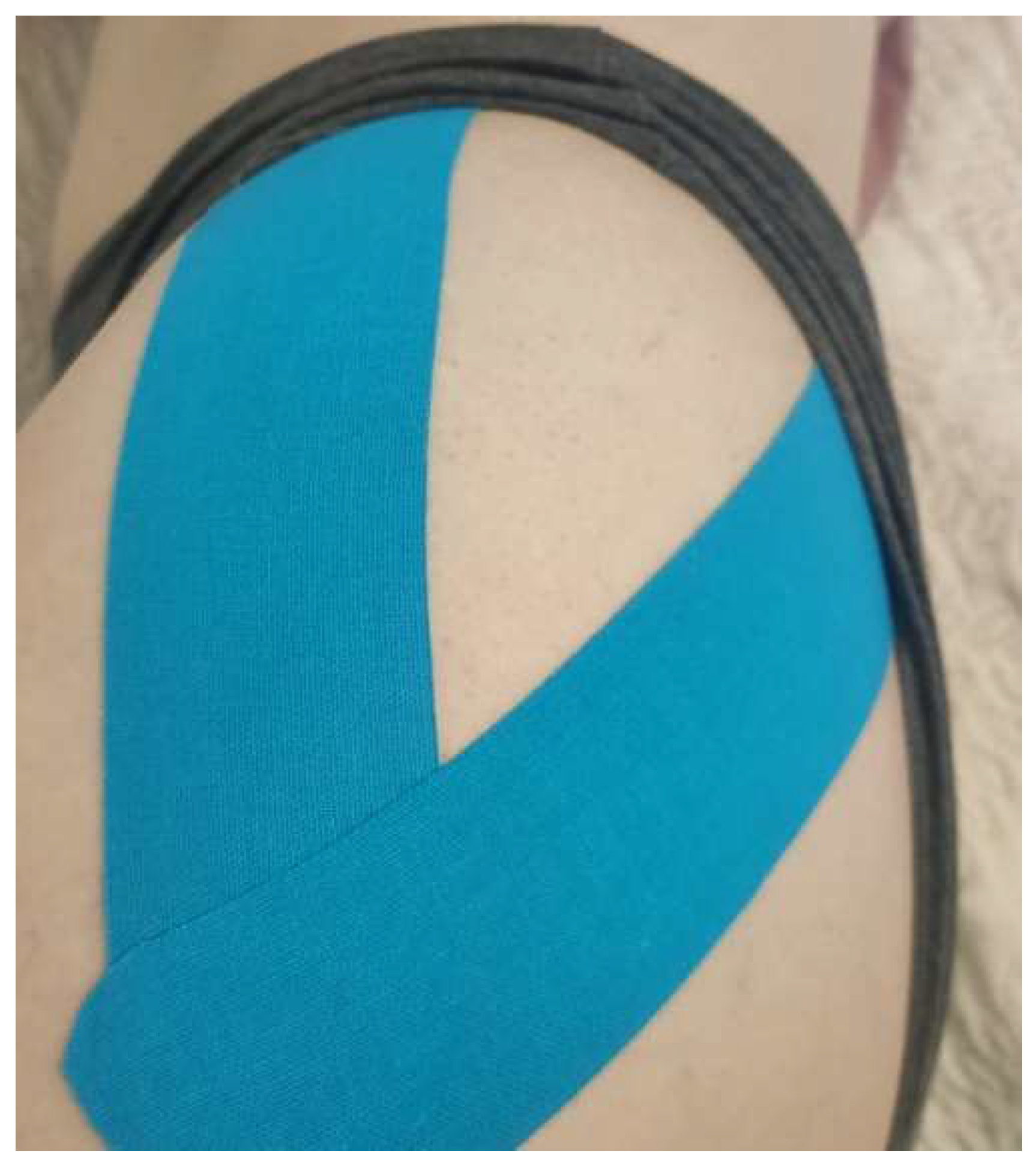
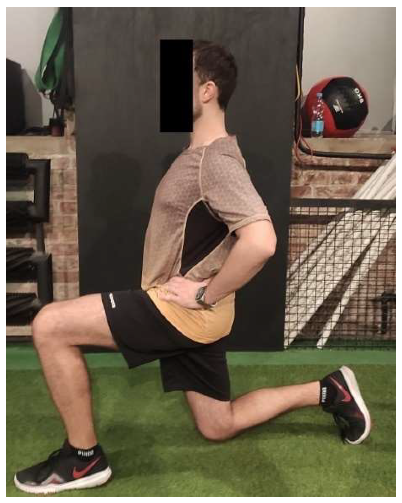
2.3. Procedures
2.4. Electromyographic Measurements
2.5. Data Analyses
3. Results
4. Discussion
Limitations
5. Conclusions
Author Contributions
Funding
Institutional Review Board Statement
Informed Consent Statement
Data Availability Statement
Conflicts of Interest
Appendix A


References
- Neumann, D.A. Kinesiology of the Hip: A Focus on Muscular Actions. J. Orthop. Sports Phys. Ther. 2010, 40, 82–94. [Google Scholar] [CrossRef] [PubMed] [Green Version]
- Presswood, L.; Cronin, J.; Keogh, J.W.L.; Whatman, C. Gluteus Medius: Applied Anatomy, Dysfunction, Assessment, and Progressive Strengthening. Strength Cond. J. 2008, 30, 41–53. [Google Scholar] [CrossRef]
- Nelson-Wong, E.; Gregory, D.E.; Winter, D.A.; Callaghan, J.P. Gluteus medius muscle activation patterns as a predictor of low back pain during standing. Clin. Biomech. 2008, 23, 545–553. [Google Scholar] [CrossRef] [PubMed]
- Zacharias, A.; Green, R.A.; Semciw, A.; English, D.J.; Kapakoulakis, T.; Pizzari, T. Atrophy of hip abductor muscles is related to clinical severity in a hip osteoarthritis population. Clin. Anat. 2018, 31, 507–513. [Google Scholar] [CrossRef]
- Boren, K.; Conrey, C.; Le Coguic, J.; Paprocki, L.; Voight, M.; Robinson, T.K. Electromyographic analysis of gluteus medius and gluteus maximus during rehabilitation exercises. Int. J. Sports Phys. Ther. 2011, 6, 206–223. [Google Scholar]
- Sheng, Y.; Duan, Z.; Qu, Q.; Chen, W.; Yu, B. Kinesio taping in treatment of chronic non-specific low back pain: A systematic review and meta-analysis. J. Rehabil. Med. 2019, 51, 734–740. [Google Scholar] [CrossRef] [Green Version]
- Lu, Z.; Li, X.; Chen, R.; Guo, C. Kinesio taping improves pain and function in patients with knee osteoarthritis: A meta-analysis of randomized controlled trials. Int. J. Surg. 2018, 59, 27–35. [Google Scholar] [CrossRef]
- Wang, Y.; Gu, Y.; Chen, J.; Luo, W.; He, W.; Han, Z.; Tian, J. Kinesio taping is superior to other taping methods in ankle functional performance improvement: A systematic review and meta-analysis. Clin. Rehabil. 2018, 32, 1472–1481. [Google Scholar] [CrossRef]
- Ghozy, S.; Dung, N.M.; Morra, M.E.; Morsy, S.; Elsayed, G.G.; Tran, L.; Minh, L.H.N.; Abbas, A.S.; Loc, T.T.H.; Hieu, T.H.; et al. Efficacy of kinesio taping in treatment of shoulder pain and disability: A systematic review and meta-analysis of randomised controlled trials. Physiotherapy 2020, 107, 176–188. [Google Scholar] [CrossRef]
- Kase, K.; Wallis, J.; Kase, T. Clinical Therapeutic Applications of the Kinesio Taping Method, 2nd ed.; Kinesio Taping Association: Albuquerque, NM, USA, 2003. [Google Scholar]
- Garnett, R.; Stephens, J.A. Changes in the recruitment threshold of motor units produced by cutaneous stimulation in man. J. Physiol. 1981, 311, 463–473. [Google Scholar] [CrossRef]
- Snodgrass, S.J.; Farrell, S.F.; Tsao, H.; Osmotherly, P.G.; Rivett, D.A.; Chipchase, L.S.; Schabrun, S.M. Shoulder Taping and Neuromuscular Control. J. Athl. Train. 2018, 53, 395–403. [Google Scholar] [CrossRef] [PubMed]
- Alahmari, K.A.; Reddy, R.S.; Tedla, J.S.; Samuel, P.S.; Kakaraparthi, V.N.; Rengaramanujam, K.; Ahmed, I. The effect of Kinesio taping on cervical proprioception in athletes with mechanical neck pain—a placebo-controlled trial. BMC Musculoskelet. Disord. 2020, 21, 648. [Google Scholar] [CrossRef] [PubMed]
- Ye, W.; Jia, C.; Jiang, J.; Liang, Q.; He, C. Effectiveness of Elastic Taping in Patients with Knee Osteoarthritis: A Systematic Review and Meta-Analysis. Am. J. Phys. Med. Rehabil. 2020, 99, 495–503. [Google Scholar] [CrossRef] [PubMed]
- Sawkins, K.; Refshauge, K.; Kilbreath, S.; Raymond, J. The placebo effect of ankle taping in ankle instability. Med. Sci. Sports Exerc. 2007, 39, 781–787. [Google Scholar] [CrossRef] [PubMed] [Green Version]
- Luz Júnior, M.A.; Sousa, M.V.; Neves, L.A.; Cezar, A.A.; Costa, L.O. Kinesio Taping® is not better than placebo in reducing pain and disability in patients with chronic non-specific low back pain: A randomized controlled trial. Braz. J. Phys. Ther. 2015, 19, 482–490. [Google Scholar] [CrossRef] [Green Version]
- Vanti, C.; Bertozzi, L.; Gardenghi, I.; Turoni, F.; Guccione, A.A.; Pillastrini, P. Effect of taping on spinal pain and disability: Systematic review and meta-analysis of randomized trials. Phys. Ther. 2015, 95, 493–506. [Google Scholar] [CrossRef] [PubMed]
- Mak, D.N.; Au, I.P.; Chan, M.; An, W.W.; Zhang, J.H.; Draper, D.; Cheung, R.T. Placebo effect of facilitatory Kinesio tape on muscle activity and muscle strength. Physiother. Theory Pract. 2019, 35, 157–162. [Google Scholar] [CrossRef]
- Parreira Pdo, C.; Costa Lda, C.; Hespanhol, L.C., Jr.; Lopes, A.D.; Costa, L.O. Current evidence does not support the use of Kinesio Taping in clinical practice: A systematic review. J. Physiother. 2014, 60, 31–39. [Google Scholar] [CrossRef] [Green Version]
- Halseth, T.; McChesney, J.W.; Debeliso, M.; Vaughn, R.; Lien, J. The effects of kinesio™ taping on proprioception at the ankle. J. Sports Sci. Med. 2004, 3, 1–7. [Google Scholar]
- Desjardins-Charbonneau, A.; Roy, J.S.; Dionne, C.E.; Desmeules, F. The Efficacy of Taping for Rotator Cuff Tendinopathy: A Systematic Review and Meta-Analysis. Int. J. Sports Phys. Ther. 2015, 10, 420–433. Available online: http://www.ncbi.nlm.nih.gov/pmc/articles/pmc4527190/ (accessed on 2 July 2022).
- Reneker, J.C.; Latham, L.; McGlawn, R.; Reneker, M.R. Effectiveness of kinesiology tape on sports performance abilities in athletes: A systematic review. Phys. Ther. Sport 2018, 31, 83–98. [Google Scholar] [CrossRef] [PubMed]
- Ramírez-Vélez, R.; Hormazábal-Aguayo, I.; Izquierdo, M.; González-Ruíz, K.; Correa-Bautista, J.E.; García-Hermoso, A. Effects of kinesio taping alone versus sham taping in individuals with musculoskeletal conditions after intervention for at least one week: A systematic review and meta-analysis. Physiotherapy 2019, 105, 412–420. [Google Scholar] [CrossRef] [PubMed]
- Warden, S.J.; Hinman, R.S.; Watson, M.A., Jr.; Avin, K.G.; Bialocerkowski, A.E.; Crossley, K. Patellar taping and bracing for the treatment of chronic knee pain: A systematic review and meta-analysis. Arthritis Care Res. 2007, 59, 73–83. [Google Scholar] [CrossRef] [PubMed]
- de Oliveira, F.C.L.; de Fontenay, B.P.; Bouyer, L.J.; Desmeules, F.; Roy, J.S. Kinesiotaping for the Rehabilitation of Rotator Cuff-Related Shoulder Pain: A Randomized Clinical Trial. Sports Health 2021, 13, 161–172. [Google Scholar] [CrossRef]
- Saracoglu, I.; Emuk, Y.; Taspinar, F. Does taping in addition to physiotherapy improve the outcomes in subacromial impingement syndrome? A systematic review. Physiother. Theory Pract. 2018, 34, 251–263. [Google Scholar] [CrossRef]
- Peterson, L.; Renstrom, P. Braces and taping used in sport. In Sports Injuries: Prevention, Treatment and Rehabilitation, 4th ed.; CRC Press: Boca Raton, FL, USA, 2017; pp. 61–63. [Google Scholar]
- Lau, K.K.; Cheng, K.C. Effectiveness of taping on functional performance in elite athletes: A systematic review. J. Biomech. 2019, 90, 16–23. [Google Scholar] [CrossRef]
- Chen, S.M.; Lo, S.K.; Cook, J. The effect of rigid taping with tension on mechanical displacement of the skin and change in pain perception. J. Sci. Med. Sport 2018, 21, 342–346. [Google Scholar] [CrossRef]
- Macgregor, K.; Gerlach, S.; Mellor, R.; Hodges, P.W. Cutaneous stimulation from patella tape causes a differential increase in vasti muscle activity in people with patellofemoral pain. J. Orthop. Res. 2005, 23, 351–358. [Google Scholar] [CrossRef]
- Alexander, C.M.; Stynes, S.; Thomas, A.; Lewis, J.; Harrison, P.J. Does tape facilitate or inhibit the lower fibres of trapezius? Man. Ther. 2003, 8, 37–41. [Google Scholar] [CrossRef]
- Mahmoud, W.S.; Kame, E.M.; Atteya, M.R.; Ibrahim, A.A.; Alrawaili, S.M.; Hussein, H.M. Immediate and short-term effects of 35% tension Kinesio tapingon handgrip strength in healthy females. Physiother. Q. 2021, 29, 9–13. [Google Scholar] [CrossRef]
- Miller, J.; Westrick, R.; Diebal, A.; Marks, C.; Gerber, J.P. Immediate effects of lumbopelvic manipulation and lateral gluteal kinesio taping on unilateral patellofemoral pain syndrome: A pilot study. Sports Health 2013, 5, 214–219. [Google Scholar] [CrossRef] [PubMed] [Green Version]
- Silva, R.O.; Carlos, F.R.; Morales, M.C.; Emerick, V.S.; Teruyu, A.I.; Valadão, V.M.A.; Carvalho, L.C.; Lobato, D.F.M. Effect of two Dynamic Tape™ applications on the electromyographic activity of the gluteus medius and functional performance in women: A randomized, controlled, clinical trial. J. Bodyw. Mov. Ther. 2021, 25, 212–217. [Google Scholar] [CrossRef] [PubMed]
- Lawrenson, P.R.; Crossley, K.M.; Vicenzino, B.T.; Hodges, P.W.; James, G.; Croft, K.J.; King, M.G.; Semciw, A.I. Muscle size and composition in people with articular hip pathology: A systematic review with meta-analysis. Osteoarthr. Cartil. 2019, 27, 181–195. [Google Scholar] [CrossRef] [PubMed]
- Bolgla, L.A.; Uhl, T.L. Electromyographic analysis of hip rehabilitation exercises in a group of healthy subjects. J. Orthop. Sports Phys. Ther. 2005, 35, 487–494. [Google Scholar] [CrossRef] [PubMed] [Green Version]
- O’Sullivan, K.; Smith, S.M.; Sainsbury, D. Electromyographic analysis of the three subdivisions of gluteus medius during weight-bearing exercises. Sports Med. Arthrosc. Rehabil. Ther. Technol. 2010, 2, 17. [Google Scholar] [CrossRef] [PubMed] [Green Version]
- Noh, K.H.; Kang, M.H.; An, S.J.; Kim, M.H.; Yoo, W.G.; Oh, J.S. Effect of Hip Joint Position on Hip Abductor Muscle Activity during Lateral Step-up Exercises. J. Phys. Ther. Sci. 2012, 24, 1145–1148. [Google Scholar] [CrossRef] [Green Version]
- Powers, C.M. The influence of altered lower-extremity kinematics on patellofemoral joint dysfunction: A theoretical perspective. J. Orthop. Sports Phys. Ther. 2003, 33, 639–646. [Google Scholar] [CrossRef]
- Kim, J.H.; Chung, Y.; Kim, Y.; Hwang, S. Functional electrical stimulation applied to gluteus medius and tibialis anterior corresponding gait cycle for stroke. Gait Posture 2012, 36, 65–67. [Google Scholar] [CrossRef]
- Baffa, A.P.; Felicio, L.R.; Saad, M.C.; Nogueira-Barbosa, M.H.; Santos, A.C.; Bevilaqua-Grossi, D. Quantitative MRI of Vastus Medialis, Vastus Lateralis and Gluteus Medius Muscle Workload after Squat Exercise: Comparison Between Squatting with Hip Adduction and Hip Abduction. J. Hum. Kinet. 2012, 33, 5–14. [Google Scholar] [CrossRef]
- Felicio, L.R.; de Carvalho, C.A.M.; Dias, C.L.C.A.; Vigário, P.D.S. Electromyographic activity of the quadriceps and gluteus medius muscles during/different straight leg raise and squat exercises in women with patellofemoral pain syndrome. J. Electromyogr. Kinesiol. 2019, 48, 17–23. [Google Scholar] [CrossRef]
- Krause, D.A.; Jacobs, R.S.; Pilger, K.E.; Sather, B.R.; Sibunka, S.P.; Hollman, J.H. Electromyographic Analysis of the Gluteus Medius in Five Weight-Bearing Exercises. J. Strength Cond. Res. 2009, 23, 2689–2694. [Google Scholar] [CrossRef] [PubMed]
- Reiman, M.P.; Bolgla, L.A.; Lorenz, D. Hip Function’s Influence on Knee Dysfunction: A Proximal Link to a Distal Problem. J. Sport Rehabil. 2009, 18, 33–46. [Google Scholar] [CrossRef] [PubMed]
- Moore, D.; Semciw, A.I.; Pizzari, T. A systematic review and meta-analysis of common therapeutic exercises that generate highest muscle activity in the gluteus medius and gluteus minimus segments. Int. J. Sports Phys. Ther. 2020, 15, 856–881. [Google Scholar] [CrossRef] [PubMed]
- Reiman, M.P.; Bolgla, L.A.; Loudon, J.K. A literature review of studies evaluating gluteus maximus and gluteus medius activation during rehabilitation exercises. Physiother. Theory Pract. 2012, 28, 257–268. [Google Scholar] [CrossRef] [PubMed]
- Alexander, C.M.; McMullan, M.; Harrison, P.J. What is the effect of taping along or across a muscle on motoneurone excitability? A study using triceps surae. Man. Ther. 2008, 13, 57–62. [Google Scholar] [CrossRef]
- Konishi, Y. Tactile stimulation with kinesiology tape alleviates muscle weakness attributable to attenuation of Ia afferents. J. Sci. Med. Sport 2013, 16, 45–48. [Google Scholar] [CrossRef]
- de Freitas, F.S.; Brown, L.E.; Gomes, W.A.; Behm, D.G.; Marchetti, P.H. No effect of kinesiology tape on passive tension, strength or quadriceps muscle activation of during maximal voluntary isometric contractions in resistance trained men. Int. J. Sports Phys. Ther. 2018, 13, 661–667. [Google Scholar] [CrossRef] [Green Version]
- Halski, T.; Dymarek, R.; Ptaszkowski, K.; Słupska, L.; Rajfur, K.; Rajfur, J.; Pasternok, M.; Smykla, A.; Taradaj, J. Kinesiology Taping does not Modify Electromyographic Activity or Muscle Flexibility of Quadriceps Femoris Muscle: A Randomized, Placebo-Controlled Pilot Study in Healthy Volleyball Players. Med. Sci. Monit. 2015, 21, 2232–2239. [Google Scholar] [CrossRef] [Green Version]
- Serrão, J.C.; Mezêncio, B.; Claudino, J.G.; Soncin, R.; Miyashiro, P.L.; Sousa, E.P.; Borges, E.; Zanetti, V.; Phillip, I.; Mochizuki, L.; et al. Effect of 3 Different Applications of Kinesio Taping Denko® on Electromyographic Activity: Inhibition or Facilitation of the Quadriceps of Males During Squat Exercise. J. Sports Sci. Med. 2016, 15, 403–409. [Google Scholar]
- Glória, I.P.D.S.; Politti, F.; Leal, E.C.P., Jr.; Lucareli, P.R.G.; Herpich, C.M.; Antonialli, F.C.; Gomes, C.A.F.D.P.; Gonzalez, T.D.O.; Biasotto-Gonzalez, D.A. Kinesio taping does not alter muscle torque, muscle activity or jumping performance in professional soccer players: A randomized, placebo-controlled, blind, clinical trial. J. Back Musculoskelet. Rehabil. 2017, 30, 869–877. [Google Scholar] [CrossRef]
- Cai, C.; Au, I.P.; An, W.; Cheung, R.T. Facilitatory and inhibitory effects of Kinesio tape: Fact or fad? J. Sci. Med. Sport 2016, 19, 109–112. [Google Scholar] [CrossRef] [PubMed]
- Au, I.P.H.; Fan, P.C.P.; Lee, W.Y.; Leong, M.W.; Tang, O.Y.; An, W.W.; Cheung, R.T. Effects of Kinesio tape in individuals with lateral epicondylitis: A deceptive crossover trial. Physiother. Theory Pract. 2017, 33, 914–919. [Google Scholar] [CrossRef] [PubMed]
- Lins, C.A.D.A.; Neto, F.L.; de Amorim, A.B.C.; Macedo, L.; Brasileiro, J.S. Kinesio Taping® does not alter neuromuscular performance of femoral quadriceps or lower limb function in healthy subjects: Randomized, blind, controlled, clinical trial. Man. Ther. 2013, 18, 41–45. [Google Scholar] [CrossRef] [PubMed]
- Lee, C.R.; Lee, D.Y.; Jeong, H.S.; Lee, M.H. The effects of Kinesiotaping on VMO and VL EMG activities during stair ascent and descent by persons with patellofemoral pain: A preliminary study. J. Phys. Ther. Sci. 2012, 24, 153–156. [Google Scholar] [CrossRef]
- Ataullah, M.G.; Kapoor, G.; Alghadir, A.H.; Khan, M. Effects of kinesio taping on hip abductor muscle strength and electromyography activity in athletes with chronic ankle instability: A randomized controlled trial. J. Rehabil. Med. 2021, 53, 2801. [Google Scholar] [CrossRef] [PubMed]
- Hsu, Y.H.; Chen, W.Y.; Lin, H.C.; Wang, W.T.; Shih, Y.F. The effects of taping on scapular kinematics and muscle performance in baseball players with shoulder impingement syndrome. J. Electromyogr. Kinesiol. 2009, 19, 1092–1099. [Google Scholar] [CrossRef]
- Słupik, A.; Dwornik, M.; Białoszewski, D.; Zych, E. Effect of Kinesio Taping on bioelectrical activity of vastus medialis muscle. Preliminary report. Ortop. Traumatol. Rehabil. 2007, 9, 644–651. [Google Scholar]
- Huang, C.Y.; Hsieh, T.H.; Lu, S.C.; Su, F.C. Effect of the Kinesio tape to muscle activity and vertical jump performance in healthy inactive people. Biomed. Eng. Online 2011, 10, 70. [Google Scholar] [CrossRef] [Green Version]
- Briem, K.; Eythörsdöttir, H.; Magnúsdóttir, R.G.; Pálmarsson, R.; Rúnarsdöttir, T.; Sveinsson, T. Effects of kinesio tape compared with nonelastic sports tape and the untaped ankle during a sudden inversion perturbation in male athletes. J. Orthop. Sports Phys. Ther. 2011, 41, 328–335. [Google Scholar] [CrossRef]
- Aydin, N.S.; Dilbay, N.K.; Selçuk, H.; Özer, A.Y. Muscle activation of the upper trapezius and functional typing performance during computer typing task: A comparison of two different wrist immobilization methods. J. Bodyw. Mov. Ther. 2021, 27, 472–476. [Google Scholar] [CrossRef]
- Cowan, S.M.; Bennell, K.L.; Hodges, P.W. Therapeutic patellar taping changes the timing of vasti muscle activation in people with patellofemoral pain syndrome. Clin. J. Sport Med. 2002, 12, 339–347. [Google Scholar] [CrossRef] [PubMed]
- Wu, C.K.; Lin, Y.C.; Lai, C.P.; Wang, H.P.; Hsieh, T.H. Dynamic Taping Improves Landing Biomechanics in Young Volleyball Athletes. Int. J. Environ. Res. Public Health 2022, 19, 13716. [Google Scholar] [CrossRef] [PubMed]
- de Sire, A.; Demeco, A.; Marotta, N.; Spanò, R.; Curci, C.; Farì, G.; Fortunato, F.; Iona, T.; Lippi, L.; Paolucci, T.; et al. Neuromuscular Impairment of Knee Stabilizer Muscles in a COVID-19 Cluster of Female Volleyball Players: Which Role for Rehabilitation in the Post-COVID-19 Return-to-Play? Appl. Sci. 2022, 12, 557. [Google Scholar] [CrossRef]
- Kim, H.H.; Kim, K.H. Effects of Kinesio Taping with Squat Exercise on the Muscle Activity, Muscle Strength, Muscle Tension, and Dynamic Stability of Softball Players in the Lower Extremities: A Randomized Controlled Study. Int. J. Environ. Res. Public Health 2021, 19, 276. [Google Scholar] [CrossRef] [PubMed]


| Group | Total N (%) | V Kramer | Chi2 | p | |||
|---|---|---|---|---|---|---|---|
| Kinesio Tape N (%) | Rigid Tape N (%) | Placebo N (%) | |||||
| Female | 18 (60) | 18 (60) | 14 (46.7) | 50 (55.6) | 0.126 | 1.440 | 0.487 |
| Male | 12 (40) | 12 (40) | 16 (53.3) | 40 (44.4) | |||
| Total | 30 (100) | 30 (100) | 30 (100) | 90 (100) | |||
| Variable | Group | N | (±SD) | Standard Error | 95% Confidence Interval | F | p | |
|---|---|---|---|---|---|---|---|---|
| Min | Max | |||||||
| Age | KT | 30 | 21.97 (0.89) | 0.16 | 21.63 | 22.30 | 0.950 | 0.391 |
| RT | 30 | 21.77 (0.94) | 0.17 | 21.42 | 22.12 | |||
| C | 30 | 21.63 (1.00) | 0.18 | 21.26 | 22.01 | |||
| Body height | KT | 30 | 169.77 (8.52) | 1.55 | 166.59 | 172.95 | 0.313 | 0.732 |
| RT | 30 | 168.57 (10.09) | 1.84 | 164.80 | 172.33 | |||
| C | 30 | 170.47 (9.54) | 1.74 | 166.91 | 174.03 | |||
| Body mass | KT | 30 | 66.83 (11.22) | 2.05 | 62.65 | 71.02 | 0.287 | 0.751 |
| RT | 30 | 66.53 (12.49) | 2.28 | 61.87 | 71.20 | |||
| C | 30 | 68.67 (11.69) | 2.13 | 64.30 | 73.03 | |||
| Body Mass Index | KT | 30 | 23.07 (2.46) | 0.45 | 22.16 | 23.99 | 0.185 | 0.832 |
| RT | 30 | 23.26 (2.79) | 0.51 | 22.22 | 24.30 | |||
| C | 30 | 23.46 (2.04) | 0.37 | 22.70 | 24.22 | |||
| Dependent Variable | Mean Difference (I-J) | Standard Error | Significance | 95% Confidence Interval | |||
|---|---|---|---|---|---|---|---|
| Min | Max | ||||||
| Glute Bridge before taping | KT | RT | −0.08 | 4.60 | 1.00 | −11.31 | 11.16 |
| C | −8.49 | 4.60 | 0.21 | −19.72 | 2.74 | ||
| RT | KT | 0.08 | 4.60 | 1.00 | −11.16 | 11.31 | |
| C | −8.41 | 4.60 | 0.21 | −19.65 | 2.82 | ||
| C | KT | 8.49 | 4.60 | 0.21 | −2.74 | 19.72 | |
| RT | 8.41 | 4.60 | 0.21 | −2.82 | 19.65 | ||
| Glute Bridge immediately after taping | KT | RT | −0.31 | 4.64 | 1.00 | −11.64 | 11.02 |
| C | −9.77 | 4.64 | 0.11 | −21.10 | 1.56 | ||
| RT | KT | 0.31 | 4.64 | 1.00 | −11.02 | 11.64 | |
| C | −9.46 | 4.64 | 0.13 | −20.79 | 1.87 | ||
| C | KT | 9.77 | 4.64 | 0.11 | −1.56 | 21.10 | |
| RT | 9.46 | 4.64 | 0.13 | −1.87 | 20.79 | ||
| Glute Bridge 48 h after taping | KT | RT | −0.04 | 4.65 | 1.00 | −11.40 | 11.32 |
| C | −8.88 | 4.65 | 0.18 | −20.24 | 2.48 | ||
| RT | KT | 0.04 | 4.65 | 1.00 | −11.32 | 11.40 | |
| C | −8.84 | 4.65 | 0.18 | −20.20 | 2.52 | ||
| C | KT | 8.88 | 4.65 | 0.18 | −2.48 | 20.24 | |
| RT | 8.84 | 4.65 | 0.18 | −2.52 | 20.20 | ||
| Unilateral Glute Bridge before taping | KT | RT | 0.36 | 5.23 | 1.00 | −12.42 | 13.13 |
| K | −9.16 | 5.23 | 0.25 | −21.93 | 3.62 | ||
| RT | KT | −0.36 | 5.23 | 1.00 | −13.13 | 12.42 | |
| C | −9.51 | 5.23 | 0.22 | −22.29 | 3.26 | ||
| C | KT | 9.16 | 5.23 | 0.25 | −3.62 | 21.93 | |
| RT | 9.51 | 5.23 | 0.22 | −3.26 | 22.29 | ||
| Unilateral Glute Bridge immediately after taping | KT | RT | −0.05 | 5.12 | 1.00 | −12.54 | 12.44 |
| C | −10.55 | 5.12 | 0.13 | −23.04 | 1.94 | ||
| RT | KT | 0.05 | 5.12 | 1.00 | −12.44 | 12.54 | |
| C | −10.50 | 5.12 | 0.13 | −22.99 | 1.99 | ||
| C | KT | 10.55 | 5.12 | 0.13 | −1.94 | 23.04 | |
| RT | 10.50 | 5.12 | 0.13 | −1.99 | 22.99 | ||
| Unilateral Glute Bridge 48 h after taping | KT | RT | 1.93 | 5.17 | 1.00 | −10.70 | 14.56 |
| C | −8.57 | 5.17 | 0.30 | −21.20 | 4.06 | ||
| RT | KT | −1.93 | 5.17 | 1.00 | −14.56 | 10.70 | |
| C | −10.50 | 5.17 | 0.14 | −23.13 | 2.13 | ||
| C | KT | 8.57 | 5.17 | 0.30 | −4.06 | 21.20 | |
| RT | 10.50 | 5.17 | 0.14 | −2.13 | 23.13 | ||
| Clamshell before taping | KT | RT | 3.44 | 5.31 | 1.00 | −9.52 | 16.41 |
| C | −4.42 | 5.31 | 1.00 | −17.38 | 8.54 | ||
| RT | KT | −3.44 | 5.31 | 1.00 | −16.41 | 9.52 | |
| C | −7.86 | 5.31 | 0.43 | −20.83 | 5.10 | ||
| C | KT | 4.42 | 5.31 | 1.00 | −8.54 | 17.38 | |
| RT | 7.86 | 5.31 | 0.43 | −5.10 | 20.83 | ||
| Clamshell immediately after taping | KT | RT | 3.13 | 5.37 | 1.00 | −9.97 | 16.23 |
| C | −7.36 | 5.37 | 0.52 | −20.46 | 5.74 | ||
| RT | KT | −3.13 | 5.37 | 1.00 | −16.23 | 9.97 | |
| C | −10.49 | 5.37 | 0.16 | −23.59 | 2.61 | ||
| C | KT | 7.36 | 5.37 | 0.52 | −5.74 | 20.46 | |
| RT | 10.49 | 5.37 | 0.16 | −2.61 | 23.59 | ||
| Clamshell 48 h after taping | KT | RT | 3.06 | 5.22 | 1.00 | −9.69 | 15.81 |
| C | −6.91 | 5.22 | 0.57 | −19.66 | 5.83 | ||
| RT | KT | −3.06 | 5.22 | 1.00 | −15.81 | 9.69 | |
| C | −9.97 | 5.22 | 0.18 | −22.72 | 2.77 | ||
| C | KT | 6.91 | 5.22 | 0.57 | −5.83 | 19.66 | |
| RT | 9.97 | 5.22 | 0.18 | −2.77 | 22.72 | ||
| Lunge before taping | KT | RT | −0.34 | 4.89 | 1.00 | −12.27 | 11.59 |
| C | 3.81 | 4.89 | 1.00 | −8.12 | 15.74 | ||
| RT | KT | 0.34 | 4.89 | 1.00 | −11.59 | 12.27 | |
| C | 4.14 | 4.89 | 1.00 | −7.79 | 16.07 | ||
| C | KT | −3.81 | 4.89 | 1.00 | −15.74 | 8.12 | |
| RT | −4.14 | 4.89 | 1.00 | −16.07 | 7.79 | ||
| Lunge immediately after taping | KT | RT | −1.29 | 4.52 | 1.00 | −12.33 | 9.75 |
| C | 0.99 | 4.52 | 1.00 | −10.05 | 12.03 | ||
| RT | KT | 1.29 | 4.52 | 1.00 | −9.75 | 12.33 | |
| C | 2.29 | 4.52 | 1.00 | −8.75 | 13.33 | ||
| C | KT | −0.99 | 4.52 | 1.00 | −12.03 | 10.05 | |
| RT | −2.29 | 4.52 | 1.00 | −13.33 | 8.75 | ||
| Lunge 48 h after taping | KT | RT | 0.00 | 4.33 | 1.00 | −10.57 | 10.57 |
| C | 5.03 | 4.33 | 0.74 | −5.53 | 15.60 | ||
| RT | KT | 0.00 | 4.33 | 1.00 | −10.57 | 10.57 | |
| C | 5.03 | 4.33 | 0.74 | −5.53 | 15.60 | ||
| C | KT | −5.03 | 4.33 | 0.74 | −15.60 | 5.53 | |
| RT | −5.03 | 4.33 | 0.74 | −15.60 | 5.53 | ||
| Pelvic Drop before taping | KT | RT | 4.24 | 4.59 | 1.00 | −6.96 | 15.43 |
| C | −6.14 | 4.59 | 0.55 | −17.34 | 5.05 | ||
| RT | KT | −4.24 | 4.59 | 1.00 | −15.43 | 6.96 | |
| C | −10.38 | 4.59 | 0.08 | −21.58 | 0.82 | ||
| C | KT | 6.14 | 4.59 | 0.55 | −5.05 | 17.34 | |
| RT | 10.38 | 4.59 | 0.08 | −0.82 | 21.58 | ||
| Pelvic Drop immediately after taping | KT | RT | 4.56 | 4.64 | 0.98 | −6.76 | 15.89 |
| C | −8.01 | 4.64 | 0.26 | −19.33 | 3.31 | ||
| RT | KT | −4.56 | 4.64 | 0.98 | −15.89 | 6.76 | |
| C | −12.57 | 4.64 | 0.02 | −23.90 | −1.25 | ||
| C | KT | 8.01 | 4.64 | 0.26 | −3.31 | 19.33 | |
| RT | 12.57 | 4.64 | 0.02 | 1.25 | 23.90 | ||
| Pelvic Drop 48 h after taping | KT | RT | 5.34 | 4.57 | 0.74 | −5.80 | 16.48 |
| C | −5.48 | 4.57 | 0.70 | −16.62 | 5.67 | ||
| RT | KT | −5.34 | 4.57 | 0.74 | −16.48 | 5.80 | |
| C | −10.82 | 4.57 | 0.06 | −21.96 | 0.33 | ||
| C | KT | 5.48 | 4.57 | 0.70 | −5.67 | 16.62 | |
| RT | 10.82 | 4.57 | 0.06 | −0.33 | 21.96 | ||
| Test | Group | I—Before Taping | II—Immediately After Taping | I vs. II | III—48 h After Taping | I vs. III | |||||||||||
|---|---|---|---|---|---|---|---|---|---|---|---|---|---|---|---|---|---|
| (±SD) | Mean Standard Error | Min | Max | (±SD) | Mean Standard Error | Min | Max | t | p | (±SD) | Mean Standard Error | Min | Max | t | p | ||
| Glute Bridge | KT | 33.50 (16.19) | 2.96 | 27.45 | 39.54 | 36.47 (15.38) | 2.81 | 30.73 | 42.22 | −3.902 | 0.001 * | 35.14 (15.83) | 2.89 | 29.23 | 41.05 | −2.226 | 0.034 * |
| RT | 33.57 (18.39) | 3.36 | 26.71 | 40.44 | 36.78 (18.34) | 3.35 | 29.94 | 43.63 | −5.098 | 0.001 * | 35.18 (18.39) | 3.36 | 28.32 | 42.05 | −2.153 | 0.040 * | |
| C | 41.99 (18.79) | 3.43 | 34.97 | 49.00 | 46.24 (19.91) | 3.63 | 38.81 | 53.67 | −5.872 | 0.001 * | 44.03 (19.64) | 3.59 | 36.69 | 51.36 | −1.324 | 0.196 | |
| Σ | 36.35 (18.07) | 1.91 | 32.57 | 40.14 | 39.83 (18.35) | 1.93 | 35.99 | 43.67 | −2.87 | 0.005 * | 38.12 (18.31) | 1.93 | 34.28 | 41.95 | −2.87 | 0.005 * | |
| Unilateral Glute Bridge | KT | 45.09 (20.49) | 3.74 | 37.43 | 52.74 | 48.08 (19.84) | 3.62 | 40.67 | 55.48 | −2.422 | 0.022 * | 47.56 (20.51) | 3.74 | 39.90 | 55.22 | −1.670 | 0.106 |
| RT | 44.73 (20.42) | 3.73 | 37.11 | 52.35 | 48.13 (19.53) | 3.56 | 40.84 | 55.42 | −2.374 | 0.024 * | 45.63 (20.06) | 3.66 | 38.14 | 53.12 | −0.558 | 0.581 | |
| C | 54.24 (19.87) | 3.63 | 46.82 | 61.66 | 58.63 (20.10) | 3.67 | 51.12 | 66.13 | −4.728 | 0.001 * | 56.13 (19.53) | 3.57 | 48.84 | 63.43 | −1.878 | 0.071 | |
| Σ | 48.02 (20.52) | 2.16 | 43.72 | 52.32 | 51.61 (20.22) | 2.13 | 47.37 | 55.85 | −5.15 | 0.001 * | 49.78 (20.34) | 2.14 | 45.52 | 54.03 | −2.20 | 0.030 * | |
| Clamshell | KT | 53.79 (21.69) | 3.96 | 45.69 | 61.89 | 56.11 (22.24) | 4.06 | 47.81 | 64.42 | −2.741 | 0.010 * | 54.90 (22.00) | 4.02 | 46.68 | 63.11 | −1.429 | 0.164 |
| RT | 50.35 (18.64) | 3.40 | 43.39 | 57.31 | 52.98 (19.93) | 3.64 | 45.54 | 60.42 | −2.994 | 0.006 * | 51.84 (19.78) | 3.61 | 44.45 | 59.22 | −1.086 | 0.287 | |
| C | 58.21 (21.23) | 3.88 | 50.28 | 66.14 | 63.47 (20.11) | 3.67 | 55.96 | 70.98 | −5.384 | 0.001 * | 61.81 (18.75) | 3.42 | 54.81 | 68.81 | −2.141 | 0.061 | |
| Σ | 54.12 (20.59) | 2.17 | 49.80 | 58.43 | 57.52 (21.02) | 2.22 | 53.12 | 61.93 | −6.38 | 0.001 * | 56.18 (20.43) | 2.15 | 51.90 | 60.46 | −2.69 | 0.009 * | |
| Pelvic Drop | KT | 41.99 (17.81) | 3.25 | 35.34 | 48.64 | 44.52 (18.35) | 3.35 | 37.66 | 51.37 | −4.044 | 0.001 * | 44.31 (17.69) | 3.23 | 37.70 | 50.92 | −3.485 | 0.002 * |
| RT | 37.75 (15.89) | 2.90 | 31.82 | 43.69 | 39.95 (16.12) | 2.94 | 33.94 | 45.97 | −3.012 | 0.005 * | 38.97 (15.98) | 2.92 | 33.00 | 44.94 | −1.443 | 0.160 | |
| C | 48.13 (19.42) | 3.55 | 40.88 | 55.39 | 52.53 (19.27) | 3.52 | 45.33 | 59.72 | −5.875 | 0.001 * | 49.79 (19.22) | 3.51 | 42.61 | 56.96 | −1.529 | 0.137 | |
| Σ | 42.63 (18.08) | 1.91 | 38.84 | 46.41 | 45.67 (18.51) | 1.95 | 41.79 | 49.54 | −7.34 | 0.001 * | 44.36 (18.04) | 1.90 | 40.58 | 48.13 | −3.43 | 0.001 * | |
| Lunge | KT | 44.09 (20.67) | 3.77 | 36.37 | 51.81 | 46.39 (19.84) | 3.62 | 38.98 | 53.80 | 2.015 | 0.011 * | 46.16 (19.81) | 3.62 | 38.76 | 53.55 | −1.303 | 0.203 |
| RT | 44.43 (18.73) | 3.42 | 37.44 | 51.42 | 47.68 (17.15) | 3.13 | 41.28 | 54.09 | −1.787 | 0.084 | 46.16 (16.90) | 3.09 | 39.85 | 52.47 | −1.379 | 0.179 | |
| C | 40.29 (17.23) | 3.15 | 33.85 | 46.72 | 45.40 (15.24) | 2.78 | 39.71 | 51.09 | 45.40 | 0.001 * | 41.12 (12.86) | 2.35 | 36.32 | 45.92 | −0.510 | 0.614 | |
| Σ | 42.94 (18.81) | 1.98 | 39.00 | 46.88 | 46.49 (17.34) | 1.83 | 42.86 | 50.12 | −4.07 | 0.001 * | 44.48 (16.75) | 1.77 | 40.97 | 47.99 | −1.80 | 0.076 | |
Publisher’s Note: MDPI stays neutral with regard to jurisdictional claims in published maps and institutional affiliations. |
© 2022 by the authors. Licensee MDPI, Basel, Switzerland. This article is an open access article distributed under the terms and conditions of the Creative Commons Attribution (CC BY) license (https://creativecommons.org/licenses/by/4.0/).
Share and Cite
Zaworski, K.; Baj-Korpak, J.; Kręgiel-Rosiak, A.; Gawlik, K. Effects of Kinesio Taping and Rigid Taping on Gluteus Medius Muscle Activation in Healthy Individuals: A Randomized Controlled Study. Int. J. Environ. Res. Public Health 2022, 19, 14889. https://doi.org/10.3390/ijerph192214889
Zaworski K, Baj-Korpak J, Kręgiel-Rosiak A, Gawlik K. Effects of Kinesio Taping and Rigid Taping on Gluteus Medius Muscle Activation in Healthy Individuals: A Randomized Controlled Study. International Journal of Environmental Research and Public Health. 2022; 19(22):14889. https://doi.org/10.3390/ijerph192214889
Chicago/Turabian StyleZaworski, Kamil, Joanna Baj-Korpak, Anna Kręgiel-Rosiak, and Krystyna Gawlik. 2022. "Effects of Kinesio Taping and Rigid Taping on Gluteus Medius Muscle Activation in Healthy Individuals: A Randomized Controlled Study" International Journal of Environmental Research and Public Health 19, no. 22: 14889. https://doi.org/10.3390/ijerph192214889