DE69831752T2 - PROCESS FOR THE PREPARATION OF L-DIHYDROOROTIC ACID AND ITS USE - Google Patents
PROCESS FOR THE PREPARATION OF L-DIHYDROOROTIC ACID AND ITS USE Download PDFInfo
- Publication number
- DE69831752T2 DE69831752T2 DE69831752T DE69831752T DE69831752T2 DE 69831752 T2 DE69831752 T2 DE 69831752T2 DE 69831752 T DE69831752 T DE 69831752T DE 69831752 T DE69831752 T DE 69831752T DE 69831752 T2 DE69831752 T2 DE 69831752T2
- Authority
- DE
- Germany
- Prior art keywords
- dho
- serum
- dihydroorotic acid
- cells
- mpa
- Prior art date
- Legal status (The legal status is an assumption and is not a legal conclusion. Google has not performed a legal analysis and makes no representation as to the accuracy of the status listed.)
- Expired - Fee Related
Links
Classifications
-
- C—CHEMISTRY; METALLURGY
- C07—ORGANIC CHEMISTRY
- C07D—HETEROCYCLIC COMPOUNDS
- C07D239/00—Heterocyclic compounds containing 1,3-diazine or hydrogenated 1,3-diazine rings
- C07D239/02—Heterocyclic compounds containing 1,3-diazine or hydrogenated 1,3-diazine rings not condensed with other rings
- C07D239/20—Heterocyclic compounds containing 1,3-diazine or hydrogenated 1,3-diazine rings not condensed with other rings having two double bonds between ring members or between ring members and non-ring members
- C07D239/22—Heterocyclic compounds containing 1,3-diazine or hydrogenated 1,3-diazine rings not condensed with other rings having two double bonds between ring members or between ring members and non-ring members with hetero atoms directly attached to ring carbon atoms
-
- G—PHYSICS
- G01—MEASURING; TESTING
- G01N—INVESTIGATING OR ANALYSING MATERIALS BY DETERMINING THEIR CHEMICAL OR PHYSICAL PROPERTIES
- G01N30/00—Investigating or analysing materials by separation into components using adsorption, absorption or similar phenomena or using ion-exchange, e.g. chromatography or field flow fractionation
- G01N30/02—Column chromatography
-
- B—PERFORMING OPERATIONS; TRANSPORTING
- B01—PHYSICAL OR CHEMICAL PROCESSES OR APPARATUS IN GENERAL
- B01J—CHEMICAL OR PHYSICAL PROCESSES, e.g. CATALYSIS OR COLLOID CHEMISTRY; THEIR RELEVANT APPARATUS
- B01J41/00—Anion exchange; Use of material as anion exchangers; Treatment of material for improving the anion exchange properties
- B01J41/20—Anion exchangers for chromatographic processes
-
- G—PHYSICS
- G01—MEASURING; TESTING
- G01N—INVESTIGATING OR ANALYSING MATERIALS BY DETERMINING THEIR CHEMICAL OR PHYSICAL PROPERTIES
- G01N30/00—Investigating or analysing materials by separation into components using adsorption, absorption or similar phenomena or using ion-exchange, e.g. chromatography or field flow fractionation
- G01N30/02—Column chromatography
- G01N30/88—Integrated analysis systems specially adapted therefor, not covered by a single one of the groups G01N30/04 - G01N30/86
- G01N2030/8809—Integrated analysis systems specially adapted therefor, not covered by a single one of the groups G01N30/04 - G01N30/86 analysis specially adapted for the sample
- G01N2030/8813—Integrated analysis systems specially adapted therefor, not covered by a single one of the groups G01N30/04 - G01N30/86 analysis specially adapted for the sample biological materials
- G01N2030/8831—Integrated analysis systems specially adapted therefor, not covered by a single one of the groups G01N30/04 - G01N30/86 analysis specially adapted for the sample biological materials involving peptides or proteins
-
- G—PHYSICS
- G01—MEASURING; TESTING
- G01N—INVESTIGATING OR ANALYSING MATERIALS BY DETERMINING THEIR CHEMICAL OR PHYSICAL PROPERTIES
- G01N30/00—Investigating or analysing materials by separation into components using adsorption, absorption or similar phenomena or using ion-exchange, e.g. chromatography or field flow fractionation
- G01N30/02—Column chromatography
- G01N30/26—Conditioning of the fluid carrier; Flow patterns
- G01N30/28—Control of physical parameters of the fluid carrier
- G01N30/32—Control of physical parameters of the fluid carrier of pressure or speed
Description
Die Erfindung betrifft ein Verfahren zur Gewinnung von L-Dihydroorotsäure (im Nachstehenden „L-DHO" bezeichnet) durch Chromatographie an einem anionischen Austauschmaterial in einem Basen-Wasser-Gemisch unter einem Druck von 1,1 MPa bis 40 MPa. Das Verfahren kann verwendet werden, um die in-vitro- und in-vivo-Aktivität von N-(4-Trifluormethylphenyl)-5-methylisoxazol-4-carboxamid, N-(4-Trifluormethylphenyl)-2-cyano-3-hydroxycrotonamid und ähnliche Verbindungen zu untersuchen.The The invention relates to a process for the recovery of L-dihydroorotic acid (in Hereinafter referred to as "L-DHO") Chromatography on an anionic exchange material in one Base-water mixture under a pressure of 1.1 MPa to 40 MPa. The Method can be used to assess the in vitro and in vivo activity of N- (4-trifluoromethylphenyl) -5-methylisoxazole-4-carboxamide, N- (4-trifluoromethylphenyl) -2-cyano-3-hydroxycrotonamide and similar To investigate connections.
L-DHO kann durch ein Kieselgel-Chromatographieverfahren mit anschließender chemischer Derivatisierung und colorimetrischer Bestimmung (Kesner, L., Aronson, F.L., Silverman, M., Chan, P.C., Clin. Chem 21/3 (1975) 353) bestimmt werden. Ein weiteres Verfahren wandelt L-DHO enzymatisch zu Orotsäure durch L-Dihydroorotsäuredehydrogenase (im Nachstehenden „DHODH"), hergestellt aus Rattenleber, um und weist nach chemischer Derivatisierung Orotat durch colorimetrische Veränderungen nach (Rogers, L.E., Nicolaisen, K., Experientia 28/10 (1972) 1259). Die Nachteile von diesen Verfahren sind die Störung von anderen Materialien in komplexen physiologischen Lösungen. Außerdem sind die erwähnten Verfahren auf Grund von arbeitsaufwändiger Probenherstellung sehr zeitraubend und deshalb für Routineanalysen in großen klinischen Studien nicht anwendbar.L-DHO can by a silica gel chromatography process followed by chemical Derivatization and colorimetric determination (Kesner, L., Aronson, F.L., Silverman, M., Chan, P.C., Clin. Chem 21/3 (1975) 353) become. Another method enzymatically converts L-DHO to orotic acid by L-dihydroorotic acid dehydrogenase (Hereinafter "DHODH") made of Rat liver, and shows after chemical derivatization orotate by colorimetric changes according to (Rogers, L.E., Nicolaisen, K., Experientia 28/10 (1972) 1259). The disadvantages of these methods are the interference of other materials in complex physiological solutions. Furthermore are the mentioned ones Procedure due to laborious sample preparation very time consuming and therefore for Routine analysis in large clinical studies not applicable.
Bei der Bemühung zur Bereitstellung von verbesserten Trennungs- und Isolierungsverfahren zur Gewinnung von L-Dihydroorotsäure wurde nun gefunden, dass dieselben durch Chromatographie von L-DHO in einem Basen-Wasser-Gemisch an einem anionischen Austauschmaterial und bei einem Druck von 1,1 MPa bis 40 MPa erzielt werden können. Das Verfahren kann für die quantitative Bestimmung von L-DHO in Zelllysaten, Säugerserum und Humanserum verwendet werden. Das Verfahren ist sehr reproduzierbar, empfindlich und verlässlich.at the effort to provide improved separation and isolation processes for the recovery of L-dihydroorotic acid has now been found to be the same by chromatography of L-DHO in a base-water mixture on an anionic exchange material and can be achieved at a pressure of 1.1 MPa to 40 MPa. The Procedure can for the quantitative determination of L-DHO in cell lysates, mammalian serum and human serum. The process is very reproducible, sensitive and reliable.
Die Erfindung, wie in den Ansprüchen erläutert, löst die Aufgabe durch ein chromatographisches Verfahren, umfassend die Schritte von:
- a) Bereitstellen einer Säule, umfassend druckstabiles anionisches Austauschmaterial;
- b) Beladen der Säule mit einer Probenlösung, die L-Dihydroorotsäure einschließt;
- c) Ausführen von Chromatographie;
- d) Eluieren der L-Dihydroorotsäure mit einer eluierenden Lösung, enthaltend ein Basen-Wasser-Gemisch;
- a) providing a column comprising pressure-stable anionic exchange material;
- b) loading the column with a sample solution including L-dihydroorotic acid;
- c) performing chromatography;
- d) eluting the L-dihydroorotic acid with an eluting solution containing a base-water mixture;
Der Begriff druckstabiles anionisches Austauschmaterial bedeutet beispielsweise Materialien, wie makroporöses (2 000 Å) Divinylbenzol/Ethylvinylbenzolpolymer oder ein mikroporöses Polyvinylbenzylammoniumpolymer, vernetzt mit Divinylbenzol oder Gemischen davon, die mit Alkanol-quaternärem Ammonium modifiziert sind; oder Vinylbenzylchlorid/Divinylbenzol-makroporöses Polymer; oder vernetztes Polyethyliminopolymer; oder Siliziumdioxid, modifiziert mit Propyltrimethylammonium; oder Poly(styrol-divinylbenzol)trimethylammonium.Of the Term pressure-stable anionic exchange material means, for example Materials, such as macroporous (2,000 Å) Divinylbenzene / ethylvinylbenzene polymer or a microporous polyvinylbenzylammonium polymer, crosslinked with divinylbenzene or mixtures thereof with alkanol quaternary ammonium are modified; or vinylbenzyl chloride / divinylbenzene macroporous polymer; or crosslinked polyethyleneimine copolymer; or silica, modified with propyltrimethylammonium; or poly (styrene-divinylbenzene) trimethylammonium.
Die
nachstehenden Produkte sind besonders bevorzugt:
Ion Pac AS
11, CarboPac PA 1 oder CarboPac MA 1 Anionenaustauschsäulen, bezogen
von Dionex Corporation, Idstein, Deutschland,
GROM – SIL, starkes
Anion, oder GROM – SIL,
schwaches Anion; bezogen von Grom P 1000 SAX, Ionospher SA oder
Chrompack PA; bezogen von Chrompack PRP-X100 oder RCX-10, bezogen
von Hamilton.The following products are particularly preferred:
Ion Pac AS 11, CarboPac PA 1 or CarboPac MA 1 anion exchange columns purchased from Dionex Corporation, Idstein, Germany,
GROM - SIL, strong anion, or GROM - SIL, weak anion; purchased from Grom P 1000 SAX, Ionospher SA or Chrompack PA; purchased from Chrompack PRP-X100 or RCX-10, supplied by Hamilton.
Die Elutionslösung enthält ein Basen-Wasser-Gemisch. Geeignete Basen sind abgeleitet von Alkalimetallen oder Erdalkalimetallen, wie Natriumhydroxid, Kaliumhydroxid, Magnesiumhydroxid oder Calciumhydroxid. Die Konzentration der Base ist 1 mMol/l bis etwa 200 mMol/l, bezogen auf Wasser als Lösungsmittel, vorzugsweise 2 mMol/l bis etwa 120 mMol/l, besonders bevorzugt ist 100 mMol/l.The elution contains a base-water mixture. Suitable bases are derived from alkali metals or alkaline earth metals, such as sodium hydroxide, potassium hydroxide, magnesium hydroxide or calcium hydroxide. The concentration of the base is 1 mmol / l to about 200 mmol / l, based on water as solvent, preferably 2 from mmol / l to about 120 mmol / l, more preferably 100 mmol / l.
Die Temperatur während des Chromatographieverfahrens ist etwa 0°C bis etwa 50°C, vorzugsweise etwa 15°C bis etwa 30°C, besonders bevorzugt etwa 19°C bis etwa 25°C. Der Arbeitsdruck während der Chromatographie ist im Wesentlichen konstant. Die Chromatographie kann unter Verwendung von verschiedenen Drücken ausgeführt werden; beispielsweise kann die Chromatographie unter einem Druck von 1,1 × 10⁸ Pa (1,1 MPa) bis 40 × 106 Pa (40 MPa), insbesondere von 4,1 MPa bis 5,5 MPa, ausgeführt werden. Die Elutionsmittelfließgeschwindigkeiten sind von etwa 0,2 ml/min bis etwa 3 ml/min, vorzugsweise 1 ml/min.The temperature during the chromatography process is about 0 ° C to about 50 ° C, preferably about 15 ° C to about 30 ° C, more preferably about 19 ° C to about 25 ° C. The working pressure during the chromatography is essentially constant. The chromatography can be carried out using different pressures; For example, the chromatography may be carried out under a pressure of 1.1 × 10 8 Pa (1.1 MPa) to 40 × 10 6 Pa (40 MPa), especially from 4.1 MPa to 5.5 MPa. Eluant flow rates are from about 0.2 ml / min to about 3 ml / min, preferably 1 ml / min.
Die Beladung der Säulen, Chromatographie und Elution von dem L-DHO findet durch bekannte, herkömmliche technische Verfahren statt.The loading of the columns, chromatography and elution of the L-DHO is by known, forth conventional technical procedures.
Eine geeignete Elution ist jene, worin die Elution einen Zeitgradienten der Basenkonzentration, vorzugsweise mit einem linearen Verlauf, zeigt. Dieser Konzentrationsgradient kann beispielsweise durch eine niedere Basenkonzentration (null im Grenzfall), die bei der Elution am Beginn der Elution vorliegt, und durch Erhöhen der Basenkonzentration während des Elutionsverfahrens angewendet werden. Es ist auf diese Weise möglich, eine besonders wirksame Trennung von der L-DHO in Proben, abgeleitet von Serum oder Zelllysaten, zu erreichen. Ein bevorzugter Basengradient variiert von nahe 1% NaOH (100 mMol/l) und 99% Wasser (am Beginn der Elution) bis etwa 60% NaOH und 40% Wasser (am Ende der Elution), wobei der besonders bevorzugte Bereich etwa 1% NaOH und 99% Wasser (am Beginn der Elution) bis etwa 15% NaOH und 75% Wasser (am Ende der Elution) ist. Der Basen-Wasser-Gradient wird in einer li nearen Weise von 2,5 Minuten bis etwa 14 Minuten und von 14 Minuten bis etwa 25 Minuten verändert, wobei das Abfallen des Gradienten während dieser 2 Zeiträume verschieden ist.A suitable elution is that in which the elution is a time gradient the base concentration, preferably with a linear course, shows. This concentration gradient can, for example, by a lower Base concentration (zero in the limit), that in the elution at the beginning elution, and by increasing the base concentration while of the elution procedure. It is possible in this way, one particularly effective separation of the L-DHO in samples derived of serum or cell lysates. A preferred base gradient varies from near 1% NaOH (100 mmol / L) and 99% water (at the beginning elution) to about 60% NaOH and 40% water (at the end of the elution), the most preferred range is about 1% NaOH and 99% water (at the beginning of the elution) to about 15% NaOH and 75% water (at the end the elution) is. The base-water gradient is in a li nearen From 2.5 minutes to about 14 minutes and from 14 minutes to changed about 25 minutes, wherein the falling of the gradient is different during these 2 periods is.
Eine besonders geeignete Elution kann durch Anwenden einer niedrigen Basenkonzentration am Beginn des Trennverfahrens von etwa 1% für einen Zeitraum von etwa 2,5 Minuten erreicht werden. Das Ergebnis ist Eluieren von dem Meisten des störenden Materials von der biologischen Matrix von der Säule. Die Abtrennung des Analyten wird durch langsames Erhöhen des Gradienten bis etwa 23% der Base innerhalb eines Zeitraums von 14 Minuten Gesamtanalysenzeit erreicht. Dann wird die Basenkonzentration auf etwa 60% innerhalb 4 Minuten erhöht, um die Elution von stark gebundenem Material zu erlauben. 60% Base sollten für nicht länger als 6 Minuten angewendet werden, bis erneute Gleichgewichtseinstellung durch 1% Basen-Wasser-Gemisch erfolgt. Die nächste Analyse beginnt nach 45 Minuten Gesamtanalysenzeit.A Particularly suitable elution can be achieved by applying a low Base concentration at the beginning of the separation process of about 1% for a Period of about 2.5 minutes can be achieved. The result is Elute from most of the disturbing Material from the biological matrix of the column. The separation of the analyte is by slowly increasing of the gradient to about 23% of the base within a period of 14 minutes total analysis time reached. Then the base concentration becomes increased to about 60% within 4 minutes, to the elution of strong to allow bound material. 60% Base should not be longer than 6 minutes until re-equilibration by 1% base-water mixture. The next analysis starts after 45 minutes total analysis time.
Das Wasser in dem Basen-Wasser-Gradienten wurde des-ionisiert und entgast.The Water in the base water gradient was des-ionized and degassed.
Das Abtrennungsverfahren gemäß der Erfindung findet in einem Säulenverfahren statt. Die Temperatur, die vorzugsweise während der anionischen Austauschchromatographie konstant gehalten wird, kann innerhalb eines breiten Bereichs variiert werden. Ein bevorzugter Temperaturbereich ist etwa –10°C bis etwa 50°C, insbesondere etwa 15°C bis etwa 25°C.The Separation method according to the invention takes place in a column method instead of. The temperature, preferably during anionic exchange chromatography is kept constant, can vary within a wide range become. A preferred temperature range is about -10 ° C to about 50 ° C, especially about 15 ° C to about 25 ° C.
Die Elution von L-DHO findet von 10 Minuten bis 12 Minuten nach dem Beginn des Gradienten statt. Die Versuchszeit des Elutionsverfahrens ist 13 min bis 25 min. Die L-DHO wird durch einen Leitfähigkeitsdetektor, wie ein Modell CD20 von Dionex Cooperation, nachgewiesen. Um die Grundlinienverschiebung zu minimieren und die Hintergrundleitfähigkeit zu senken, kann ein Anionenselbstregenerierungssupressor, wie Modell ASRS-I, 4 mm, von Dionex Cooperation, angewendet werden.The Elution of L-DHO takes place from 10 minutes to 12 minutes after Beginning of the gradient instead. The experimental time of the elution process is 13 min to 25 min. The L-DHO is controlled by a conductivity detector, as demonstrated by a model CD20 from Dionex Cooperation. To the To minimize baseline shift and the background conductivity can lower an anion auto-regeneration suppressor, like model ASRS-I, 4 mm, from Dionex Cooperation.
Das erfindungsgemäße Verfahren ist besonders für analytische Chromatographie geeignet, kann jedoch auch für prä parative Chromatographie, insbesondere, wenn das erfindungsgemäße Verfahren mit einem präparativen Hochdruck-Flüssigchromatographiesäule-(HPLC)-System ausgeführt wird, verwendet werden. Der Begriff „präparative Chromatographie" bedeutet ein Reinigungsverfahren mit dem Ziel der Gewinnung und nicht nur Analyse von reinen Produkten. Die Menge der reinen Produkte kann innerhalb breiter Grenzen, beispielsweise 1 mg bis 1000 g, vorzugsweise zwischen 50 mg und 500 mg, variieren.The inventive method is especially for analytical chromatography, but can also be used for preparative Chromatography, especially when the inventive method with a preparative High pressure Flüssigchromatographiesäule- (HPLC) system, accomplished will be used. The term "preparative chromatography" means a purification process with the aim of obtaining and not just analyzing pure products. The amount of pure products can be within wide limits, for example 1 mg to 1000 g, preferably between 50 mg and 500 mg.
Das erfindungsgemäße Verfahren kann verwendet werden, um Veränderungen in intrazellulären oder extrazellulären L-DHO-Konzentrationen auf Grund der Inhibierung von Dihydroorotsäuredehydrogenase (DHO-DH) nachzuweisen. Das Enzym DHO-DH ist für die Umwandlung von L-Dihydroorotsäure zu Orotsäure während der De-novo-Pyrimidinsynthese verantwortlich. Die Inhibierung von DHO-DH führt zur Akkumulation von L-DHO. Das erfindungsgemäße Verfahren kann für die Herstellung eines diagnostischen Assays verwendet werden. Das erfindungsgemäße Verfahren kann zum Bestimmen der Aktivität von DHO-DH-Inhibitoren verwendet werden. DHO-DH-Inhibitoren sind beispielsweise N-4-(Trifluormethylphenyl)-5-methylisoxazol-4-carboxylamid, Brequinar, N-(4-Trifluormethylphenyl)-2-cyano-3-hydroxyhept-2-en-6-in-carboxamid, 2-Cyano-3-cyclopropyl-3-hydroxyacrylsäure-(4-cyanophenyl)-amid oder N-(4-Trifluormethylphenyl)-2-cyano-3-hydroxycrotonamid. Das erfindungsgemäße Verfahren kann zum Bestimmen von L-DHO-Konzentrationen in Pflanzen, Zelllinien, Tieren und Menschen verwendet werden. L-DHO-Bestimmung kann verwendet werden, um Aktivität von DHO-DH-Inhibitoren in Pflanzen, Säugern und Menschen zu verfolgen.The inventive method Can be used to change in intracellular or extracellular L-DHO-concentrations due to the inhibition of dihydroorotic dehydrogenase (DHO-DH). The enzyme DHO-DH is for the conversion of L-dihydroorotic acid to orotic acid during the De novo pyrimidine synthesis responsible. The inhibition of DHO-DH leads to Accumulation of L-DHO. The process according to the invention can be used for the production a diagnostic assay. The inventive method can be used to determine the activity used by DHO-DH inhibitors. DHO-DH inhibitors are for example N-4- (trifluoromethylphenyl) -5-methylisoxazole-4-carboxylamide, Brequinar, N- (4-trifluoromethylphenyl) -2-cyano-3-hydroxyhept-2-en-6-ynecarboxamide, 2-cyano-3-cyclopropyl-3-hydroxyacrylic acid (4-cyanophenyl) -amide or N- (4-trifluoromethylphenyl) -2-cyano-3-hydroxycrotonamide. The inventive method can be used to determine L-DHO concentrations in plants, cell lines, Animals and humans are used. L-DHO determination can be used to activity of DHO-DH inhibitors in plants, mammals and to persecute people.
Das erfindungsgemäße Verfahren wird genauer in den folgenden Beispielen beschrieben. Sofern nicht anders ausgewiesen, betreffen Prozentsatzdaten das Gewicht.The inventive method is described in more detail in the following examples. Unless otherwise reported, percentage data relates to the weight.
Beispiel 1example 1
1.1. Chemikalien und Reagenzien1.1. Chemicals and reagents
Chemikalien
und Reagenzien wurden wie nachstehend ausgewiesen bezogen:
Desionisiertes Wasser musste durch Helium vor der Verwendung entgast werden.deionized Water had to be degassed by helium before use.
1.2. Chromatographische Ausrüstung1.2. chromatographic equipment
Das
HPLC-System bestand aus den nachstehenden Instrumenten: 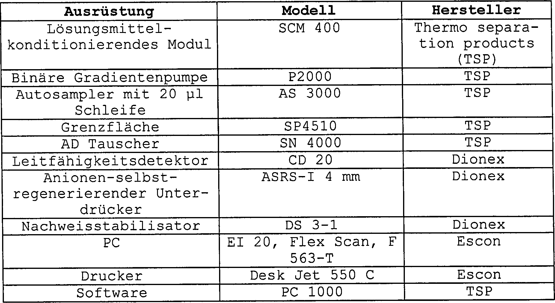
Das Peakmaterial wurde innerhalb der gesamten Versuche verwendet.The Peak material was used throughout the experiments.
1.3. HPLC-Bedingungen1.3. HPLC conditions
Die
chromatographische Abtrennung wurde mit einer 250 × 4 mm I.D.
IonPac AS 11 Anionenaustauschsäule
(Teilchengröße 13 μm; P/N 044076,
Dionex), ausgerüstet
mit einer IonPac AG 11, 50 × 4
mm I.D. Vorsäule
(Teilchengröße 13 μm; P/N 044078,
Dionex), ausgeführt.
Zusätzlich
wurde eine Anionenfallensäule ATC-1
(P/N 037151, Dionex) zwischen der Gradientenpumpe und dem Einspritzventil
installiert. Um die Grundlinienverschiebung zu minimieren und die
Hintergrundleitfähigkeit
zu senken, wurde der ASRS-I-Suppressor installiert, der bei 300
mA betrieben wurde. Der Bereich des Detektors ist mit 10 μS ausgewiesen.
Der Autosampler wurde auf 14°C
gekühlt,
jedoch die Analyse selbst wurde bei Raumtemperatur ausgeführt. Die
mobile Phase war aus 100 mM NaOH (A) und desionisiertem und entgastem
Wasser (B) zusammengesetzt. Mit dem System wurde der nachstehende
Gradient erzeugt: 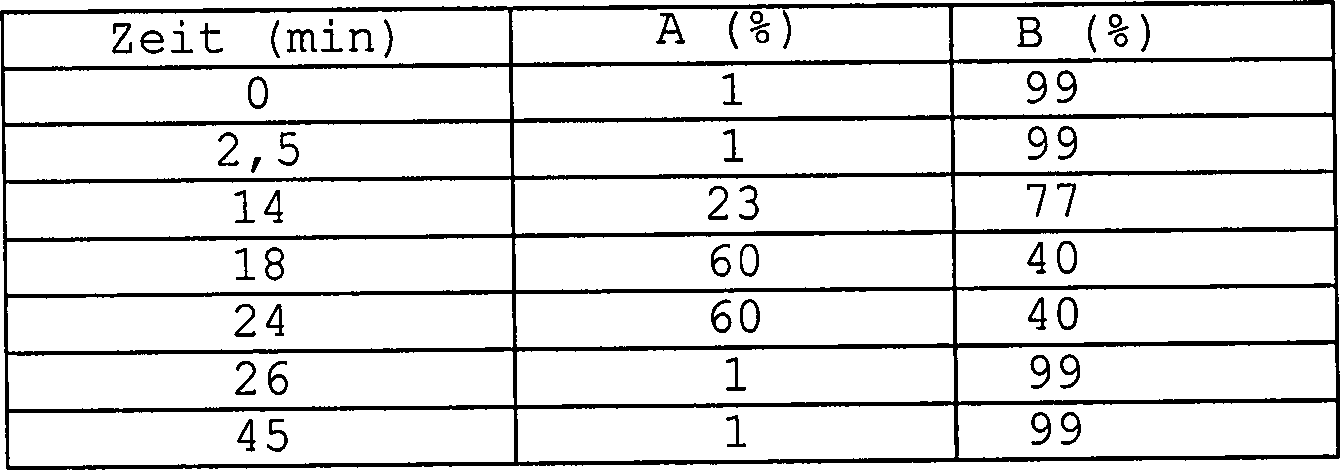
Die Fließgeschwindigkeit war 1 ml/min; Versuchszeit war 45 min.The flow rate was 1 ml / min; Trial time was 45 min.
1.4. Standards und Qualitätskontrollproben1.4. Standards and quality control samples
Eine Standardstammlösung wurde durch Auflösen von 1 mg L-DHO in 1 ml Wasser hergestellt. Aliquote Mengen von 400 μl wurden auf –20°C gefroren. Die Stabilität von diesen Lösungen wurde für mindestens 4 Wochen garantiert. Definierte Mengen der Stammlösung wurden zu den Zelllysaten und zu Human- oder Rattenserum gegeben und zum Bewerten der Linearität zwischen dem Anstieg des Signals und der festgesetzten L-DHO-Konzentration bewertet. Die Präzision und Genauigkeit des Verfahrens wurden durch Anwenden von Qualitätskontroll-(QC)-Proben mit einem L-DHO-Gehalt in dem unteren, dem mittleren und dem hohen Konzentrationsbereich der Signal/Konzentrations-Linearitätskurve untersucht.A Standard stock solution was by dissolving of 1 mg L-DHO in 1 ml of water. Aliquots of 400 μl were added frozen at -20 ° C. The stability from these solutions was for guaranteed for at least 4 weeks. Defined amounts of the stock solution were to the cell lysates and to human or rat serum and to Evaluate the linearity evaluated between the rise of the signal and the set L-DHO concentration. The precision and method accuracy were determined by applying Quality Control (QC) samples to a L-DHO content in the lower, middle and high concentration range the signal / concentration linearity curve examined.
1.5. Probenzubereitungen1.5. sample preparations
1.5.1. Zellen1.5.1. cell
Jurkat-Zellen wurden von ATCC (TIB 182) erhalten und wie in 1.5.1.1. beschrieben gezüchtet.Jurkat cells were obtained from ATCC (TIB 182) and as described in 1.5.1.1. described bred.
1.5.1.1 Gewebskulturbedingungen1.5.1.1 Tissue culture conditions
Jurkat-Zellen wurden bei 5 × 10⁵/ml angesetzt und 24 Stunden in RPMI 1640 Medium und 10%igem fötalem Kalbsserum (FCS) gezüchtet. Die Zellen wurden in frischem Medium zur Zelllyse vereinigt und die Menge der Zellen und der Prozentsatz an toten Zellen durch Vital-Mikroskopie mit Eosin berechnet. Nach 24 Stunden hatten sich Zellzahlen um 1,6–1,8-fach innerhalb weniger als 7% toter Zellen erhöht. Jurkat-Zellen, die zur L-DHO-Bestimmung genommen wurden, hatten ein Proliferationsverhältnis von 1,6–1,8-fach in 24 Stunden und weniger als 7% tote Zellen.Jurkat cells were seeded at 5 x 10 5 / ml and cultured for 24 hours in RPMI 1640 medium and 10% fetal calf serum (FCS). The cells were pooled in fresh medium for cell lysis, and the amount of cells and percentage of dead cells were calculated by vital microscopy with eosin. After 24 hours, cell numbers had increased 1.6-1.8-fold within less than 7% of dead cells. Jurkat cells taken for L-DHO determination had a proliferation ratio of 1.6-1.8 fold in 24 hours and less than 7% dead cells.
1.5.1.2 Herstellung von Zelllysaten1.5.1.2 Production of cell lysates
Zellen wurden in einem definierten Mediumvolumen suspendiert und die Zelldichte der Proben wurde über Vitalmikroskopie unter Verwendung von Eosin bestimmt. Etwa 10 × 106 Zellen wurden entfernt, mit 5 Minuten-Zentrifugation bei 350 × G pelletisiert und der Überstand wurde verworfen. Die Lyse der Zellen wurde durch Resuspendieren des Zellpellets in 500 μl 1,2M HClO4 ausgeführt. Dieses Gemisch wurde in 2,0 ml Eppendorf-Safe-Lock-Becher (Sicherheitsverschlussbecher) überführt und das Protein wurde nach 2 Minuten Hochgeschwindigkeitszentrifugierung ausgefällt. Der Überstand wurde vollständig entfernt, in Glasfläschchen überführt und nach Zusatz von 500 μl Chloroform sorgfältig vermischt durch 2 Minuten Vortexbehandlung. Zelllipide wurden durch Chloroform, gefolgt von 10 Minuten Zentrifugierung (1502 G) bei 10°C extrahiert. Der gereinigte Überstand wurde in 2 ml Eppendorf-Bechern gesammelt und bei –20°C bis zur weiteren Verwendung gelagert. Für die HPLC-Analyse wurden 100 μl dieses Überstands mit 30 μl 6M KOH neutralisiert. Nach Schütteln für etwa 5 s wurden die Proben auf Eis für etwa 30 Minuten gelagert. Anschließend wurden sie 5 min bei 15 000 U/min zentrifugiert. Von dem klaren Überstand wurden 20 μl für die HPLC-Analyse verwendet.Cells were suspended in a defined volume of medium and the cell density of the samples was determined by vital microscopy using eosin. Approximately 10 x 10 6 cells were removed, pelleted with 5 minutes centrifugation at 350 x G, and the supernatant discarded. Lysis of the cells was performed by resuspending the cell pellet in 500 ul 1.2M HClO. 4 This mixture was transferred to 2.0 ml Eppendorf Safe-Lock beaker and the protein was precipitated after 2 minutes of high speed centrifugation. The supernatant was completely removed, transferred to glass vials and, after adding 500 μl of chloroform, thoroughly mixed by vortexing for 2 minutes. Cell lipids were extracted by chloroform followed by centrifugation (1502G) for 10 minutes at 10 ° C. The purified supernatant was collected in 2 ml Eppendorf cups and stored at -20 ° C until further use. For HPLC analysis, 100 μl of this supernatant was neutralized with 30 μl of 6M KOH. After shaking for about 5 seconds, the samples were stored on ice for about 30 minutes. They were then centrifuged for 5 min at 15,000 rpm. From the clear supernatant, 20 μl was used for HPLC analysis.
1.5.2. Serum1.5.2. serum
Um den Proteingehalt zu vermindern, wurden 200 μl Serum zu einem Microconfilter (10 000 D, Modell 10, Code 42407, Amicon) gegeben und 30 min bei 13 000 Umdrehungen pro Minute (U/min) zentrifugiert. Der Durchfluss bestand aus etwa 150 μl und enthält den Analyten L-DHO. Von dieser Flüssigkeit wurden 20 μl für die HPLC-Analyse verwendet.Around To reduce the protein content, 200 μl of serum became a microconfilter (10 000 D, Model 10, Code 42407, Amicon) and 30 min at Centrifuged at 13,000 revolutions per minute (rpm). The flow consisted of about 150 μl and contains the analyte L-DHO. From this liquid was added 20 μl for HPLC analysis used.
1.6. Quanitifizierung1.6. quantitation
Der Integrator bestimmte die Peakhöhe des Analyten. Eichkurven wurden erhalten durch Auftragen der gemessenen Peakhöhen (y) gegen die Analytenkonzentration in verschiedenen biologischen Matrizes. Gewichtete lineare Regression (1/y) wurde verwendet, um die L-DHO-Konzentration in Standardproben sowie in Qualitätskontrollen zurückzurechnen. Der übliche Korrelationskoeffizient R wurde durch PROC GLM, bezogen auf die Analyse des Kovarianzmodells unter Verwendung des Wägefaktors, bereitgestellt.Of the Integrator determined the peak height of the analyte. Calibration curves were obtained by plotting the measured peak heights (y) against the analyte concentration in various biological Matrices. Weighted linear regression (1 / y) was used to the L-DHO concentration in standard samples as well as in quality controls back-calculate. The usual Correlation coefficient R was determined by PROC GLM, based on the Analysis of the covariance model using the weighing factor, provided.
1.7. Stabilität1.7. stability
Tabelle
1 zeigt die Stabilitätsdaten
des Analyten bei –20°C in Zelllysaten
und Serumproben nach 2–3 Gefrier/Auftauzyklen
von einer Probe. L-DHO war unter den vorstehend erwähnten Bedingungen
in Jurkat-Zellen über
einen Zeitraum von mindestens 4 Wochen stabil. Die nachgewiesene
Erhöhung
in der Konzentration von einigen Rattenserumproben und einer Humanserumprobe
nach einigen Auftauzyklen von mehr als 10% kann nicht erklärt werden
und zeigt, dass die Genauigkeit in solchen Fällen bis zu 15% im Allgemeinen
und bis zu 29% im schlechtesten Fall vermindert sein kann. Aus diesen
Gründen
kann nur mitgeteilt werden, dass bei Ratten- und Hu manserumproben
L-DHO letztendlich für
nur mindestens 1 Woche stabil ist. Tabelle
1: Stabilitätsdaten
in Zelllysaten und Serum bei –20°C (n = 1) 
Der
Rückstand
(%) ist ein Prozentsatz der Konzentration, verglichen mit der Anfangsanalyse.
n.d.
= nicht bestimmtThe residue (%) is a percentage of the concentration compared to the initial analysis.
nd = not determined
Um die Bedingungen der Proben zu simulieren, wenn sie in dem Autosampler auf die tatsächliche Analyse warten, wurde die Stabilität während 18 h bei 14°C bestimmt. Aus dem Grund wurden die Zelllysate versetzt und dann mit 30 μl 6M KOH/100 μl Lysat vor dem Beginn der Analyse behandelt. Entsprechend wurden die Serumproben versetzt und dann, wie in 1.5.2. beschrieben, deproteinisiert.Around to simulate the conditions of the samples when in the autosampler on the actual After waiting for analysis, the stability was determined at 14 ° C for 18 h. Therefore, the cell lysates were spiked and then pre-incubated with 30 μl 6M KOH / 100 μl lysate treated at the beginning of the analysis. Accordingly, the serum samples and then, as in 1.5.2. described, deproteinized.
Wie
in Tabelle 2 ersichtlich werden kann, ist der L-DHO-Gehalt leicht in Zellen unter diesen
Bedingungen bis zu einem Maximum von etwa 7% vermindert. In Serumproben
ist der Analyt unter diesen Bedingungen stabil. Aus diesen Gründen nur
wurden so viele HPLC-Proben hergestellt, dass die maximale Länge des
Aufenthalts in dem Autosampler unter 18 h war. Tabelle
2: Stabilität
für 18
h bei 14°C
(n = 1) 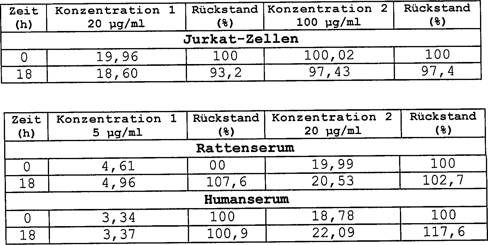
1.8. Selektivität1.8. selectivity
Der Vergleich von nicht versetzten Jurkat-Zelllysaten mit entsprechenden Chromatogrammen, unter Verwendung von Zelllysaten, die mit 50 μg L-DHO/ml versetzt wurden, zeigte, dass es einen kleinen Peak bei 11,983 min gab, was die gleiche Retentionszeit wie L-DHO ist. Es ist sehr nahe liegend, dass sich dieser Peak aus dem natürlichen Gehalt des Analyten in diesen Zellen ergibt. Zusätzlich kann ersichtlich werden, dass sich auf Grund der Behandlung der Zelllysate mit KOH der L-DHO-Peak in zwei Peaks spaltet. Die KOH-Behandlung jedoch ist wesentlich, um das saure Zelllysat vor der HPLC-Analyse zu neutralisieren. Es konnte gezeigt werden, dass unter den beschriebenen Bedingungen die Entwicklung der zweiten Peakhöhe (Retentionszeit (RT) = 11,954 min) angewendet werden kann, um verbesserte Ergebnisse bezüglich Linearität und Reproduzierbarkeit zu erhalten.Of the Comparison of Untranslated Jurkat Cell Lysates with Corresponding Chromatograms using cell lysates containing 50 μg L-DHO / ml showed that there was a small peak at 11.983 min gave what is the same retention time as L-DHO. It is very close lying, that this peak is from the natural content of the analyte in these cells. additionally can be seen that due to the treatment of the Cell lysates with KOH cleave the L-DHO peak into two peaks. The KOH treatment however, it is essential to remove the acid cell lysate prior to HPLC analysis to neutralize. It could be shown that under the described Conditions the evolution of the second peak height (retention time (RT) = 11.954 min) can be applied to improved results in terms of linearity and reproducibility to obtain.
Auch im Fall von Ratten- und Humanserum wurde ein Blindwert mit der gleichen Retentionszeit wie L-DHO gefunden. Es wird vermutet, dass sich dies auf den natürlichen L-DHO-Gehalt des Organismus bezieht. Die Bestimmung von mindestens 10 verschiedenen Proben von beiden Arten zeigte, dass der natürliche L-DHO-Gehalt unter der Nachweisgrenze von 1 μg/ml war.Also in the case of rat and human serum was a blank with the same Retention time as L-DHO found. It is believed that this is on the natural L-DHO content of Organism refers. The determination of at least 10 different Samples from both species showed that the natural L-DHO content was below that Detection limit of 1 μg / ml was.
1.9. Linearität1.9. linearity
Die Linearität der Bestimmung wurde an fünf Eichkurven für die Zelllinie und verschiedenen Serumproben bewertet. Die Proben wurden hergestellt und an fünf verschiedenen Tagen mit L-DHO-Konzentrationen im Bereich von 1,5–150 μg/ml (Zelllysate) und 1–30 μg/ml (Serumproben) laufen lassen. Die Ergebnisse werden in Tabellen 3–5 gezeigt. Für die Bestimmung der Regressionslinie wurde die Peakhöhe verwendet. Basierend auf den entsprechenden Konzentrationen der verschiedenen Standards wurde es, wie in verschiedenen Tabellen ausgewiesen, zurückberechnet.The linearity the provision was sent to five Calibration curves for evaluated the cell line and various serum samples. Samples were made and at five different days with L-DHO concentrations in the range of 1.5-150 μg / ml (cell lysates) and 1-30 μg / ml (serum samples) let run. The results are shown in Tables 3-5. For the Determination of the regression line, the peak height was used. Based on the corresponding concentrations of different standards it is recalculated as shown in various tables.
Tabelle 3 Linearität von L-DHO-Bestimmungen in Jurkat-ZelllysatenTable 3 Linearity of L-DHO determinations in Jurkat cell lysates
Nach
Versetzen wurden die Proben mit 30 μl 6M KOH behandelt und dann
einmal, wie in 1.3. beschrieben, analysiert. 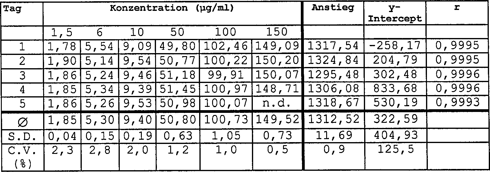
- r = 0,9995
- r = 0.9995
Tabelle 4 Linearität von L-DHO-Bestimmungen in RattenserumTable 4 Linearity of L-DHO determinations in rat serum
Nach
Versetzen wurde das Serum von den Proteinen, wie in 1.5.2. beschrieben,
gereinigt. 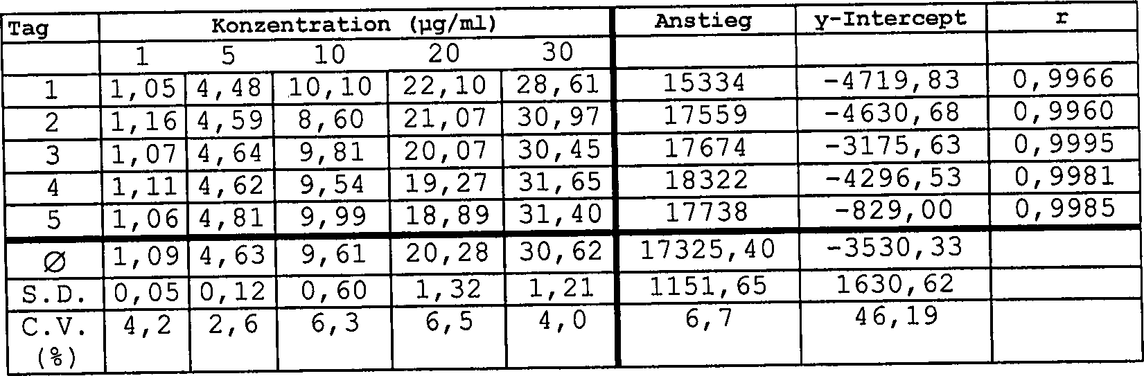
- r = 0,9959
- r = 0.9959
Tabelle 5 Linearität von L-DHO-Bestimmungen in HumanserumTable 5 Linearity of L-DHO determinations in human serum
Nach
Versetzen wurde das Serum von Proteinen, wie in 1.5.2. beschrieben,
gereinigt. 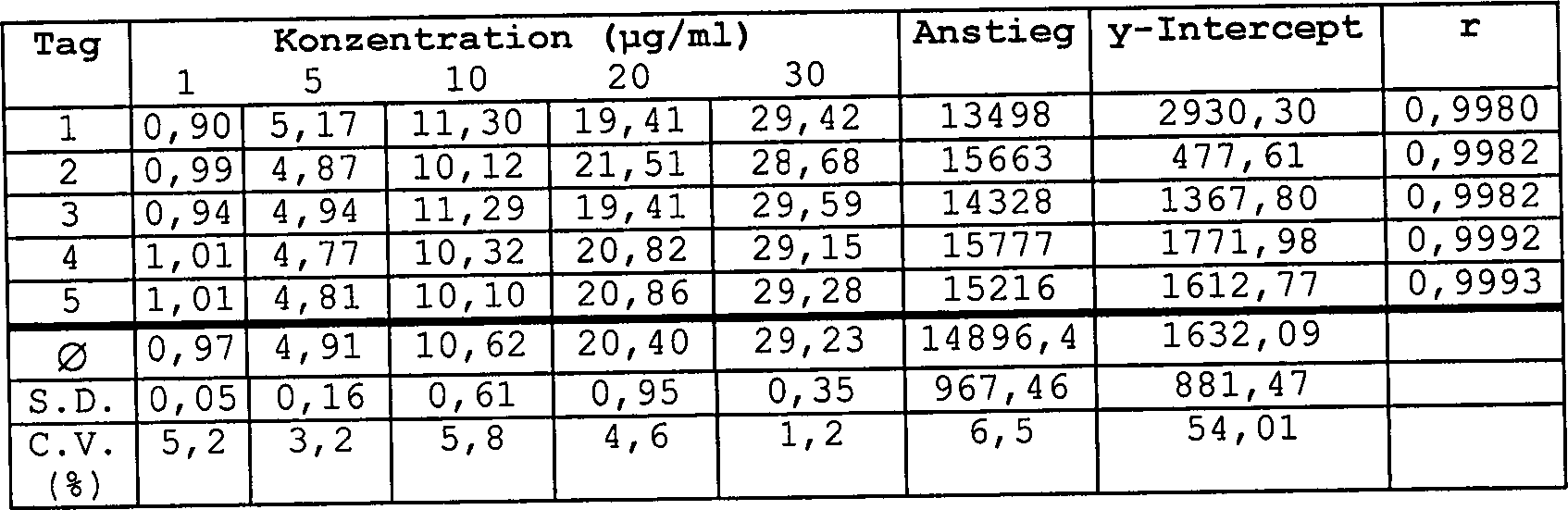
- r = 0,9986·Standardkonzentration (μg/ml) – nicht bestimmt
- r = 0.9886 · standard concentration (μg / ml) - not determined
Wie in diesen Tabellen ersichtlich werden kann, war die Linearität in jedem Fall erwiesen. Dies wird auf die jeweiligen Korrelationskoeffizienten „r" zurückgeführt, die in allen Fällen > 0,99 sind. Die mittlere lineare Regressionslinie für die an 5 verschiedenen Tagen erhaltenen Konzentrationskurven wird in jeder Tabelle beschrieben und zeigt, dass der Anstieg mit einer maximalen Variation von 6,7% sehr gut reproduzierbar war.As In these tables, the linearity was in each Case proven. This is attributed to the respective correlation coefficients "r" which in all cases> 0.99. The middle linear regression line for the concentration curves obtained on 5 different days described in each table and shows that the increase with a maximum variation of 6.7% was very well reproducible.
Die zurückgerechneten Standardkonzentrationen zeigten im Mittelwert ein C.V. von weniger als 6,5%, was die hohe Genauigkeit der Werte zeigt. Der übliche Korrelationswert r von mehr als 0,99 drückt die sehr hohe Präzision und Reproduzierbarkeit des Verfahrens aus.The back-calculated Standard concentrations averaged a C.V. less than 6.5%, which shows the high accuracy of the values. The usual correlation value r of more than 0.99 presses the very high precision and reproducibility of the method.
1.10. Quantifizierungsgrenze1.10. LOQ
Bezogen auf diese Ergebnisse, ist die Quantifizierungsgrenze in Jurkat-Zellen 1,5 μg/ml. In Ratten- und Humanserumproben kann 1 μg/ml L-DHO nachgewiesen werden. Versetzte Proben bei dieser Konzentration zeigten ein Signal zu Geräusch-Verhältnis von mindestens 1:3.Based On top of these results, the quantification limit is in Jurkat cells 1.5 μg / ml. In rat and human serum samples 1 μg / ml L-DHO can be detected. Staggered samples at this concentration indicated a signal Noise ratio of at least 1: 3.
1.12. Genauigkeit und Präzision1.12. Accuracy and precision
Genauigkeit und Präzision von wiederholten Bestimmungen von L-DHO bei drei verschiedenen Konzentrationen an fünf verschiedenen Tagen wird in Tabelle 6–8 zusammengefasst. Die Genauigkeit wird als der Prozent Unterschied von gefundener zu zugegebener Menge an L-DHO (Wiedergewinnung) ausgedrückt. Die Intra-Tag-Präzision, ausgedrückt als C.V. (%) wurde unter Verwendung der zwei Werte, die erhalten werden, wenn eine Probe zweimal am Tag gemessen wurde, berechnet. Die Inter-Tag-Präzision wurde auch wie C.V. (%) ausgedrückt und wurde unter Verwendung des Durchschnitts für Werte von jeder Kontrollprobe an 5 verschiedenen Tagen berechnet.accuracy and precision of repeated determinations of L-DHO at three different concentrations at five Different days are summarized in Table 6-8. The precision is found as the percent difference from found to added amount expressed in terms of L-DHO (recovery). The intra-day precision, expressed as C.V. (%) was obtained using the two values obtained are calculated when a sample is measured twice a day. The inter-day precision was also like C.V. (%) and was calculated using the average for values of each control calculated on 5 different days.
Tabelle 6:Table 6:
Genauigkeit und Präzision in Jurkat-Zelllysaten nach Versetzen und anschließender Neutralisation mit 30 μl 6M KOH. n = 2 bedeutet, dass eine Zelllysatprobe mit der entsprechenden Konzentration versetzt und zweimal gemessen wurde.accuracy and precision in Jurkat cell lysates after displacement and subsequent neutralization with 30 μl 6M KOH. n = 2 means that a cell lysate sample with the appropriate Concentrated and measured twice.

Tabelle 7:Table 7:
Genauigkeit und Präzision in Rattenserum nach Versetzen und anschließender Deproteinisierung; n = 2 bedeutet, dass eine Serumprobe mit der entsprechenden Konzentration versetzt und zweimal gemessen wurde.accuracy and precision in rat serum after displacement and subsequent deproteinization; n = 2 means that a serum sample with the appropriate concentration offset and measured twice.

Tabelle 8:Table 8:
Genauigkeit und Präzision in Humanserum nach Versetzen und anschließender Deproteinisierung; n = 2 bedeutet, dass eine Serumprobe mit der entsprechenden Konzentration versetzt und zweimal gemessen wurde.accuracy and precision in human serum after displacement and subsequent deproteinization; n = 2 means that a serum sample with the appropriate concentration offset and measured twice.

Die hier dargestellten Ergebnisse zeigen, dass in den meisten Fällen die Kontrollwerte ± 10% Variation bei einem Maximum aufwiesen und dass das Verfahren deshalb sehr genau ist. Nur in einem Fall unterschied sich die Wiedergewinnung in Jurkat-Zellenbestimmungen etwas von jenem Wert (–11,7%). Dieser Effekt kann durch die hohe Inter-Tag-Variation von 12% erklärt werden. Die Inter-Tag-Präzision in Jurkat-Zellen, Ratte- oder Humanserum war niedriger als 5%, 7% bzw. 10%. Die Inter-Tag-Präzision in allen untersuchten Matrizes war sehr niedrig mit einem C.V. von weniger als 6,0%. Dies zeigt, dass die erhaltenen Ergebnisse sehr stark reproduzierbar und präzise sind.The Results presented here show that in most cases the Control values ± 10% Variation at a maximum and that is why the process is very accurate. Only in one case was the recovery different in Jurkat cell determinations, something of that value (-11.7%). This effect can be explained by the high inter-day variation of 12%. The inter-day precision in Jurkat cells, rat or human serum was lower than 5%, 7% or 10%. The inter-day precision in all matrices examined was very low with a C.V. from less than 6.0%. This shows that the results obtained are very highly reproducible and precise are.
Beispiel 2Example 2
2.1. Gewebskulturbedingungen:2.1. tissue culture:
- – Herstellung von serumfreiem Medium: pulverisiertes Iscove-Medium (Biochrom) wurde in 10 Litern bidestilliertem Wasser, ergänzt mit 18,95 g NaCl, 11,43 g NaHCO3, 700 mg KCl, 10 ml 35%ige NaOH-Lösung und 0,5 ml 1M Mercaptoethanollösung (Riedel de Haen), gelöst und steril filtriert. Zu einem Liter hergestelltem Iscove-Medium wurden vor der Anwendung 32 mg Human-Holotransferrin, 1 g Rinderalbumin und 1,5 ml Lipide (Sigma) gegeben.- Preparation of serum free medium: Iscove powdered medium (Biochrom) was double distilled into 10 liters of water supplemented with 18.95 g NaCl, 11.43 g NaHCO3, 700 mg KCl, 10 ml 35% NaOH solution and 0.5 ml of 1 M mercaptoethanol solution (Riedel de Haen), dissolved and filtered sterile. To one liter of Iscove's medium was added 32 mg of human holotransferrin, 1 g of bovine albumin, and 1.5 ml of lipids (Sigma) prior to use.
- – Zellkultur: A20.2.J-Zellen wurden in serumfreiem Medium (37°C, 5% CO2) in einer Expansionskultur bei logarithmischem Zellwachstum gezüchtet. Die Zellen, die für das Assay genommen werden, hatten eine 2,2-fache Proliferationsrate in 24 h. Der Prozentsatz an toten Zellen war <8% (3).Cell culture: A20.2.J cells were grown in serum-free medium (37 ° C, 5% CO 2 ) in a logarithmic cell growth expansion culture. The cells taken for the assay had a 2.2-fold proliferation rate in 24 hours. The percentage of dead cells was <8% (3).
- – Behandlung von Zellen mit N-(4-Trifluormethyl)-2-cyano-3-hydroxy-crotonamid, hergestellt wie in EP-0 529 500, anschließend A77 1726, beschrieben. A77 1726 wurde in bidestilliertem Wasser (10 mM) gelöst und in serumfreiem Medium weiter verdünnt. Den Zellen wurde dann die geeignete Menge an A77 1726 zugegeben und bei 37°C und 5%igem CO2 inkubiert.- Treatment of cells with N- (4-trifluoromethyl) -2-cyano-3-hydroxy-crotonamide, prepared as described in EP-0 529 500, then A77 1726 described. A77 1726 was dissolved in bidistilled water (10 mM) and further diluted in serum-free medium. The appropriate amount of A77 1726 was then added to the cells and incubated at 37 ° C and 5% CO 2 .
2.2. Herstellung von Zelllysaten für DHO-Bestimmung:2.2. Production of cell lysates for DHO determination:
Hergestellte Zellen wurden in einem definierten Mediumvolumen und Zelldichte resuspendiert. In Abhängigkeit von dem erwarteten DHO-Gehalt, wurden zwischen 1–50 Millionen Zellen entfernt, pelletisiert (5 min, 350 × G) und der Überstand verworfen. Die Zellen wurden lysiert durch Zugeben von 500 μl 1,2M HClO4. Die Lysate wurden in 2 ml Eppendorf-Safe-Lock-Becher (Sicherheitsverschlussbecher) überführt und das Protein mit 2 min Hochgeschwindigkeitszentrifugierung ausgefällt. Die angesäuerten Lysate wurden vollständig entfernt, in Glasfläschchen überführt und nach Zusatz von 500 μl Chloroform sorgfältig vermischt durch 2 Minuten Vortexbehandlung. Zelllipide wurden nach 10 Minuten Kaltzentrifugierung (1502 G; 10°C) extrahiert. Die gereinigten Überstände wurden in 2 ml Eppendorf-Bechern zur Lagerung bei –20°C gesammelt bis zur Hochdruck-Flüssig-Chromatographie-(HPLC)-Bestimmung.Prepared cells were resuspended in a defined medium volume and cell density. Depending on the expected DHO content, between 1-50 million cells were removed, pelleted (5 min, 350 x G) and the supernatant discarded. The cells were lysed by adding 500 μl of 1.2M HClO 4 . The lysates were transferred to 2 ml Eppendorf Safe-Lock beakers and the protein precipitated with 2 min of high speed centrifugation. The acidified lysates were completely removed, transferred to glass vials and, after adding 500 μl of chloroform, thoroughly mixed by vortexing for 2 minutes. Cell lipids were extracted after 10 minutes of cold centrifugation (1502G, 10 ° C). The purified supernatants were collected in 2 ml Eppendorf beakers for storage at -20 ° C until high pressure liquid chromatography (HPLC) determination.
2.3. HPLC-Bestimmung von DHO:2.3. HPLC determination of DHO:
Die
chromatographische Trennung wurde wie in Beispiel 1 beschrieben
ausgeführt.
Der Bereich des Leitfähigkeitsdetektors
wurde auf 10 μS
eingestellt. Die Analyse wurde bei Raumtemperatur ausgeführt. Die mobile
Phase war aus 100 mM NaOH (A) und Wasser (B) zusammengesetzt. Mit
dem System wurde der nachstehende Gradient hergestellt: 
Die Fließgeschwindigkeit war 1 ml/min; Versuchszeit war 49 min.The flow rate was 1 ml / min; Trial time was 49 min.
2.4. Ergebnisse2.4. Results
A20.2.J-Zellen,
inkubiert mit A77 1726, hatten erhöhte Mengen an intrazellulärer DHO
(Tabellen 9–11). Die
in Tabelle 9 angeführten
Ergebnisse zeigen, dass DHO-Spiegel direkt mit der Zahl der extrahierten
Zellen korrelierten. Tabelle
9: Korrelation von intrazellulären
DHO-Konzentrationen und Anzahl der Zellen, die mit A77 1726 gezüchtet wurden 
A20.2.J-Zellen wurden mit 5 μM A77 1726 behandelt und 24 Stunden (37°C, 5% CO2) gezüchtet und dann für die DHO-Extraktion (n = 3) zubereitet.A20.2.J cells were treated with 5 μM A77 1726 and grown for 24 hours (37 ° C, 5% CO 2 ) and then prepared for DHO extraction (n = 3).
Um
die Zellkulturverfahren zu optimieren und das beste Zelle/A77 1726-Molaritätsverhältnis zu
bestimmen, wurden variierende Dichten von A20.2.J-Zellen, zusammen
mit A77 1726 (5 μM),
inkubiert. Die Zellen wurden bei verschiedenen Zeitpunkten abgezogen
und DHO-Konzentrationen bestimmt (Tabelle 10). Auf Grund der Tatsache,
dass DHO-Konzentrationen direkt mit der Menge an extrahierten Zellen
korrelierten (siehe Tabelle 9), wurden für die nachstehenden Versuche
DHO-Konzentrationen auf 10 × 106 Zellen in μg/ml extrapoliert. Der beste
lineare Anstieg des DHO-Spiegels wurde mit einer Dichte von 1 × 10⁶ Zellen/ml gefunden. Unter Anwendung dieser
Zelldichte wurde die Zeit, in Abhängigkeit von der Erhöhung der
intrazellulären DHO-Spiegel
in Zellen, die mit A77 1726 inkubiert wurden, untersucht. Nachweisbare
Mengen an DHO konnten nach 1 Stunde Inkubation, unabhängig von
der Arzneistoffkonzentration (Tabelle 11) bestimmt werden. Eine
lineare Erhöhung
wurde mit der größten Menge
an DHO, bestimmt nach 6 h (Tabelle 11), registriert. Nach diesem
Zeitraum wurde eine Sättigung
ohne weitere Erhöhung
von DHO beobachtet. Tabelle
10: Zelldichte und zeitabhängige
Erhöhung
von intrazellulären
DHO-Konzentrationen: 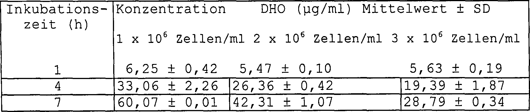
Verschiedene
Mengen an A20.2.J-Zellen wurden zusammen mit A77 1726 (5 μM) für die vorstehend angegebenen
Zeiträume
inkubiert und deren intrazelluläre
DHO-Konzentrationen für
jeden Probenpunkt bestimmt. (* Alle Werte extrapoliert auf 10 × 106 Zellen) (n = 2). Tabelle
11: Die zeitabhängige
Erhöhung
von intrazellulären
L-DHO-Konzentrationen: 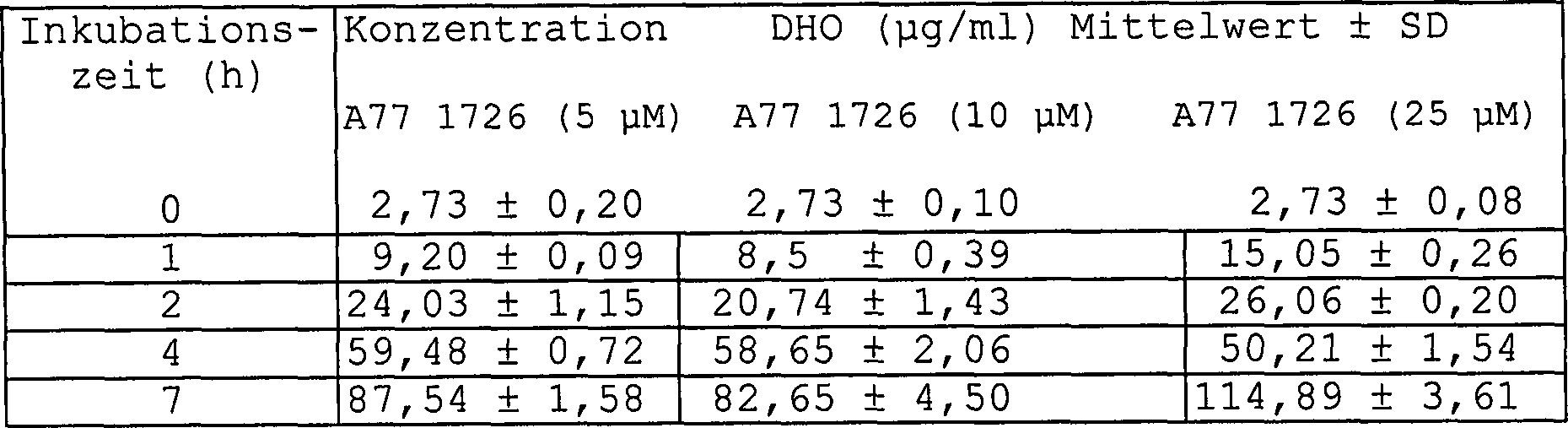
Eine Million A20.2.J-Zellen/ml wurden, zusammen mit variierenden Konzentrationen von A77 1726 und deren intrazellulären DHO-Konzentrationen, die bei verschiedenen Zeitpunkten bestimmt wurden, inkubiert. Die Daten werden als μg/ml DHO ± SD, extrapoliert auf zehn Millionen Zellen (0 – 4 h = n = 2, 7 h = n = 4), angegeben.One million A20.2.J cells / ml were added, along with varying concentrations of A77 1726 and their intracellular DHO concentrations, which were determined at different times, incubated. The data are expressed as μg / ml DHO ± SD, extrapolated to ten million cells (0-4 h = n = 2, 7 h = n = 4).
Inkubation von A20.2.J-Tumorzellen mit A77 1726 ergab schnelle Akkumulation von L-DHO auf Grund von DHO-DH-Inhibierung. Die intrazellulären L-DHO-Konzentrationen korrelierten mit der Zellzahl und waren zeitabhängig. L-DHO-Verfolgen ist ein Surrogatmarker für A77 1726 Immunomodulierungsaktivität bei Patienten.incubation of A20.2.J tumor cells with A77 1726 revealed rapid accumulation of L-DHO due to DHO-DH inhibition. The intracellular L-DHO concentrations correlated with cell number and were time dependent. L-DHO tracking is one Surrogate marker for A77 1726 Immunomodulation activity in patients.
Beispiel 3Example 3
Tiere: Männliche Wistar-Lewis-Ratten (Mollegaard Breading Center Ltd. Ejby, DK) mit einem Körpergewicht von 160–200 g.Animals: male Wistar Lewis rats (Mollegaard Breading Center Ltd Ejby, DK) with a body weight from 160-200 G.
Adjuvans Arthritis: Die Erkrankung wurde durch Injizieren von 0,1 ml Freund'schem Adjuvans (6 ml Mycobacterium smegmatis, suspendiert in 1 ml schwerem, weißem Paraffinöl (Merck, Darmstadt)), in die Schwanzwurzel von Wistar-Lewis-Ratten induziert. Pathologische Symptome treten im Allgemeinen zwischen 10 und 14 Tagen nach Krankheitsauslösung auf.adjuvant Arthritis: The disease was induced by injecting 0.1 ml of Freund's adjuvant (6 ml Mycobacterium smegmatis suspended in 1 ml white paraffin oil (Merck, Darmstadt)), into the tail root of Wistar Lewis rats. Pathological symptoms generally occur between 10 and 14 Days after illness on.
Arzneistoffbehandlung: Arzneistoffe wurden in 1%iger Carboxymethylcellulose (COMC) suspendiert. Gesunde Tiere (n = 18) und Adjuvans-erkrankte (n = 18) Ratten (Tag 9 der Störung) wurden 10 mg/kg p.o. N-4-(Trifluormethylphenyl)-5-methylisoxazol-4-carboxamid, anschließend Leflunomid zweimal täglich (7:30 und 13:30) für 5 Tage; d.h. bei Zeitpunkten: 0 h, 6 h, 24 h, 30 h, 48 h, 54 h, 72 h, 78 h, 96 h (siehe Tabelle 12), gegeben. Zum Zeitpunkt 0 Stunden erkrankten 3 von zwei Gruppen und nicht erkrankte Gruppen wurden geopfert, um die Grundlinienspiegel zu bestimmen. Weitere 3 gesunde und 3 erkrankte Tiere wurden mit Placebo (nur COMC) für 5 Tage behandelt.Drug treatment: Drugs were suspended in 1% carboxymethylcellulose (COMC). healthy Animals (n = 18) and adjuvanted (n = 18) rats (day 9 of the disorder) 10 mg / kg p.o. N-4- (trifluoromethylphenyl) -5-methylisoxazole-4-carboxamide, subsequently Leflunomide twice daily (7:30 and 13:30) for 5 days; i.e. at times: 0 h, 6 h, 24 h, 30 h, 48 h, 54 h, 72 h, 78 h, 96 h (see Table 12). At the time 0 hours Diseased 3 of two groups and non-diseased groups were sacrificed to determine the baseline levels. Another 3 healthy and 3 diseased animals were given placebo (COMC only) for 5 days treated.
Probennehmen:
Drei Tiere pro Gruppe wurden bei jedem Probennahmezeitpunkt geopfert.
Serum und Splenozyten wurden bei 3 h, 7 h, 27 h, 51 h, 75 h und
99 h (siehe Tabelle 12) genommen. Mit Ausnahme des Werts von 7 h,
wurden die Proben drei Stunden nach der letzten Arzneistoffapplikation
genommen. Der Wert 7 h wurde eine Stunde nach dem zweiten Dosieren
genommen. Proben der Placebo behandelten Tiere wurden bei den Zeitpunkten
0 h (n = 3) und 99 h (n = 3) genommen. Tabelle
12: Verabreichung von Leflunomid und Gewebsprobenahmezeitpunkte 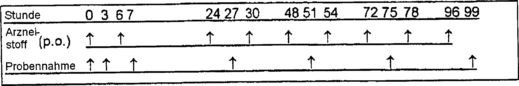
Herstellung der Proben:Preparation of samples:
- – Blut, gesammelt durch Herzpunktur, wurde 30 min bei 4°C gelagert und dann 10 min bei 3000 U/min zentrifugiert. Serum wurde abgetrennt und in Eppendorf-Bechern bei –20°C (3) gelagert. Vor HPLC-Analyse wurde das gefrorene Serum aufgetaut, und, um die Proteine zu entfernen, wurden 200 μl Serum zu einem Microconfilter (Modell 10, Code 42407, Amicon) gegeben und 30 min bei 13 000 U/min zentrifugiert.- blood, collected by cardiac puncture was stored at 4 ° C for 30 min and then at 10 min Centrifuged at 3000 rpm. Serum was separated and in Eppendorf cups stored at -20 ° C (3). Prior to HPLC analysis, the frozen serum was thawed, and to give the To remove proteins, 200 μl of serum became a Microcon filter (Model 10, code 42407, Amicon) and 30 min at 13,000 rpm centrifuged.
- – Die Milzen wurden entnommen (n = 3) und zur L-DHO-Analyse vereinigt. Die Zellen, die durch Zerlegen (Durchleiten durch ein Edelstahlsieb) getrennt wurden, wurden mit 0,17 M NH4Cl zur Lyse der Erythrozyten behandelt. Aliquote Mengen von 50 Millionen Milzzellen pro Gruppe wurden hergestellt, in Becher zur Zentrifugierung gegeben und der Überstand verworfen. Das Zellpellet wurde unter permanentem Mixen zu 500 μl einer 1,2 M HClO4-Lösung zur Lyse der Zellen gegeben und 2 min zentrifugiert. Das saure Zelllysat wurde vollständig zu Glasfläschchen überführt, 500 μl Chloroform zugesetzt und 2 min mit einem Vortexmischer vermischt. Zelluläre Lipide wurden durch Zentrifugierung (10 min 1502 × G und 10°C) ausgefällt. Der Überstand wurde in 2 ml-Becher gegeben und bei –20°C gelagert.The spleens were removed (n = 3) and pooled for L-DHO analysis. The cells separated by disassembly (passing through a stainless steel sieve) were treated with 0.17 M NH 4 Cl to lyse the erythrocytes. Aliquots of 50 million spleen cells per group were prepared, placed in beakers for centrifugation and the supernatant discarded. The cell pellet was added with permanent mixing to 500 μl of a 1.2 M HClO 4 solution to lyse the cells and centrifuged for 2 min. The acid cell lysate was completely transferred to vials, 500 μl of chloroform added and vortexed for 2 minutes. Cellular lipids were precipitated by centrifugation (10 min 1502 x G and 10 ° C). The supernatant was placed in 2 ml beakers and stored at -20 ° C.
Die
Bestimmung von A77 1726 Serumkonzentrationen wurde wie nachstehend
durchgeführt:
Serumproben
wurden auf Raumtemperatur gebracht und sorgfältig unter Anwendung eines
Vortexmischers vermischt. Das Serum wurde in Eppendorf-Becher pipettiert
(200 μl)
und der innere Standard (A77 1726, 2 μg in 400 μl Acetonitril) zugegeben. Die
Röhrchen
wurden dann in einem Vortexmischer vermischt und bei 2500 U/min
(Raumtemperatur) für
10 Minuten zentrifugiert. Zur HPLC-Analyse wurde der Überstand
(400 μl)
in ein Fläschchen überführt und
Wasser (400 μl)
zugegeben und vermischt. Die HPLC-Bedingungen waren wie nachstehend:
Die Hardware bestand aus einer TSP P2000 Pumpe, einem TSP AS1000
Autosampler, einem TSP SP4270 Integrator und einem TSP UV100 UV-Detektor.
Die Detektion war bei 292 nm Wellenlänge. Die mobile Phase bestand
aus 650 ml Methanol (CHROMASOLV), 2,42 g Tetrabutylammoniumbromid
und 350 ml 0,05 M Ammoniumacetat. Die Fließgeschwindigkeit war 0,5 ml/min.
Eine CHROMPACK Spherisorb ODS-2 Säule von 10 cm mit einer 1 cm
Umkehrphasen(R2)-Guardsäule
wurde verwendet. 100 μl
wurden in die Säule gespritzt
und die Laufzeit war 7 Minuten.The determination of A77 1726 serum concentrations was performed as follows:
Serum samples were brought to room temperature and mixed thoroughly using a vortex mixer. The serum was pipetted into Eppendorf beakers (200 μl) and the internal standard (A77 1726, 2 μg in 400 μl acetonitrile). The tubes were then vortexed and centrifuged at 2500 rpm (room temperature) for 10 minutes. For HPLC analysis, the supernatant (400 μl) was transferred to a vial and water (400 μl) added and mixed. The HPLC conditions were as follows: The hardware consisted of a TSP P2000 pump, a TSP AS1000 autosampler, a TSP SP4270 integrator, and a TSP UV100 UV detector. The detection was at 292 nm wavelength. The mobile phase consisted of 650 ml of methanol (CHROMASOLV), 2.42 g of tetrabutylammonium bromide and 350 ml of 0.05 M ammonium acetate. The flow rate was 0.5 ml / min. A 10 cm CHROMPACK Spherisorb ODS-2 column with a 1 cm reverse phase (R2) guard column was used. 100 μl was injected into the column and the runtime was 7 minutes.
HPLC-Bestimmung
von L-DHO-Konzentrationen: Die chromatographische Trennung wurde
wie in Beispiel 1 beschrieben ausgeführt. Der Bereich des Leitfähigkeitsdetektors
wurde auf 10 μS
eingestellt. Die Analyse wurde bei Raumtemperatur ausgeführt. Die
mobile Phase war aus 100 mM NaOH (A) und Wasser (B) zusammengesetzt.
Mit dem System wurden die nachstehenden Gradienten erzeugt: 
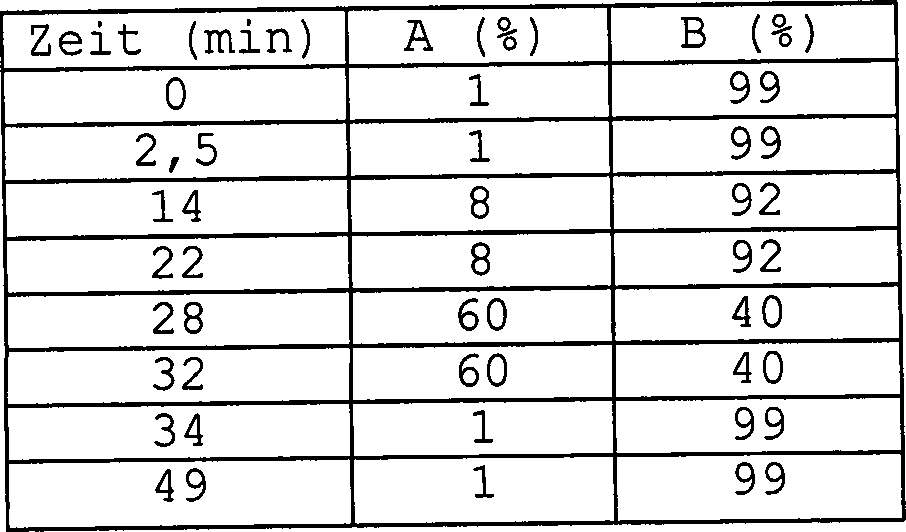
Die Fließgeschwindigkeit war 1 ml/min.The flow rate was 1 ml / min.
Sowohl
gesunde als auch erkrankte Ratten hatten erhöhte zelluläre (Tabelle 13) und Serum-(Tabelle 14)
L-DHO-Konzentrationen
nach oraler Verabreichung von Leflunomid. Diese Erhöhung korrelierte
mit den A77 1727-Serumkonzentrationen, die in diesen Tieren (Tabelle
15) bestimmt wurden. Anfänglich
erreichte 3 Stunden nach oraler Verabreichung A77 1726 eine Konzentration
von etwa 26 μg/ml
in Adjuvanserkrankten Ratten und 31 μg/ml in nicht erkrankten Ratten.
Diese Werte zeigten einen Peak eine Stunde nach dem zweiten Dosieren
(7 h), fielen jedoch auf Werte zwischen 7 und 12 μg/ml für die Dauer
des Versuchs in sowohl erkrankten als auch nicht erkrankten Ratten
ab. Erkrankte Tiere nahmen 51 h in Anspruch zum Erreichen dieser Konzentration;
wohingegen nicht erkrankte Tiere immer dieselbe nach 27 h erreichten
(Tabelle 15). Die A77 1726-Serumkonzentrationen korrelierten mit
den L-DHO-Konzentrationen in dem Serum von Adjuvanserkrankten und
nicht erkrankten Nagern. Im Gegensatz zu den L-DHO-Serumkonzentrationen,
die zu etwa 5 μg/ml
für die
Dauer des Versuchs äquilibrierten,
fiel die Menge an in den Splenozyten gefundener L-DHO unter die
Nachweisgrenze (1,5 μg/ml)
nach 99 h. Tabelle
13: L-DHO-Konzentrationen in Splenozyten [50 × 106 Zellen]
von Ratten, behandelt mit Leflunomid 
Tiere
wurden mit Leflunomid oder Placebo behandelt, die Milzen entfernt
(n = 3) und wie beschrieben vereinigt. Die vereinigten Splenozyten
wurden in zweifacher Ausführung
bestimmt. DL = Nachweisgrenze (1,5 μg/ml); Tabelle
14: L-DHO-Konzentrationen in Serum von Ratten, behandelt mit Leflunomid 
Tiere
wurden mit Leflunomid oder Placebo behandelt und Blut genommen,
präpariert
und wie beschrieben bestimmt. L-DHO-Serumkonzentrationen wurden
für jedes
Tier einzeln bestimmt. DL = Nachweisgrenze (0,5 μg/ml); Tabelle
15: A77 1726-Konzentrationen in Serum von mit Leflunomid behandelten
Ratten 
Die Tiere wurden mit Leflunomid oder Placebo behandelt und Blut genommen, präpariert und wie beschrieben bestimmt. A77 1726-Serumkonzentrationen wurden für jedes Tier einzeln bestimmt.The Animals were treated with leflunomide or placebo and taken blood, prepared and determined as described. A77 1726 serum concentrations were for each Animal determined individually.
Oral
verabreichtes Leflunomid wird sehr schnell in vivo zu A77 1726 umgewandelt.
A77 1726 ist der aktive Metabolit von Leflunomid (
Claims (10)
Applications Claiming Priority (3)
| Application Number | Priority Date | Filing Date | Title |
|---|---|---|---|
| EP97121848 | 1997-12-11 | ||
| EP97121848A EP0933633A1 (en) | 1997-12-11 | 1997-12-11 | Process for obtaining L-dihydroorotic acid and use thereof |
| PCT/EP1998/007972 WO1999030146A1 (en) | 1997-12-11 | 1998-12-08 | Process for obtaining l-dihydroorotic acid and use thereof |
Publications (2)
| Publication Number | Publication Date |
|---|---|
| DE69831752D1 DE69831752D1 (en) | 2006-02-09 |
| DE69831752T2 true DE69831752T2 (en) | 2006-06-22 |
Family
ID=8227782
Family Applications (1)
| Application Number | Title | Priority Date | Filing Date |
|---|---|---|---|
| DE69831752T Expired - Fee Related DE69831752T2 (en) | 1997-12-11 | 1998-12-08 | PROCESS FOR THE PREPARATION OF L-DIHYDROOROTIC ACID AND ITS USE |
Country Status (21)
| Country | Link |
|---|---|
| US (1) | US6545006B1 (en) |
| EP (2) | EP0933633A1 (en) |
| JP (1) | JP4189124B2 (en) |
| KR (1) | KR100700906B1 (en) |
| CN (1) | CN1237345C (en) |
| AR (1) | AR016430A1 (en) |
| AT (1) | ATE305610T1 (en) |
| AU (1) | AU747993B2 (en) |
| BR (1) | BR9813559A (en) |
| CA (1) | CA2315326A1 (en) |
| CZ (1) | CZ299968B6 (en) |
| DE (1) | DE69831752T2 (en) |
| DK (1) | DK1036319T3 (en) |
| ES (1) | ES2248927T3 (en) |
| HK (1) | HK1033171A1 (en) |
| HU (1) | HUP0004510A3 (en) |
| ID (1) | ID24807A (en) |
| PL (1) | PL194068B1 (en) |
| RU (1) | RU2228932C2 (en) |
| TR (1) | TR200001671T2 (en) |
| WO (1) | WO1999030146A1 (en) |
Families Citing this family (7)
| Publication number | Priority date | Publication date | Assignee | Title |
|---|---|---|---|---|
| KR20020067545A (en) * | 1999-12-16 | 2002-08-22 | 테바 파마슈티컬 인더스트리즈 리미티드 | Novel processes for making- and a new crystalline form of- leflunomide |
| CN104582694B (en) * | 2012-05-29 | 2018-10-02 | 线粒体科学公司研究所 | Dihydrooratic acid dehydrogenation enzyme inhibitor |
| US9679506B2 (en) | 2012-06-25 | 2017-06-13 | Sharp Kabushiki Kaisha | Multiple function display system |
| US10708575B2 (en) | 2012-06-25 | 2020-07-07 | Sharp Kabushiki Kaisha | Display system with diffuse and specular reflective modes |
| CN102787147B (en) * | 2012-08-31 | 2014-10-08 | 南京工业大学 | Method for preparing L-dihydroorotate by enzymatic method |
| US10699612B2 (en) | 2014-10-27 | 2020-06-30 | Sharp Kabushiki Kaisha | Display system with specular reflective mode |
| CN112305097A (en) * | 2020-09-30 | 2021-02-02 | 辰欣药业股份有限公司 | Method for detecting substances related to teriflunomide tablets |
Family Cites Families (3)
| Publication number | Priority date | Publication date | Assignee | Title |
|---|---|---|---|---|
| US2773872A (en) * | 1954-04-12 | 1956-12-11 | Merck & Co Inc | Dihydroorotic acid |
| HU178201B (en) * | 1977-08-05 | 1982-03-28 | Chinoin Gyogyszer Es Vegyeszet | Process for producing substituted orto-acids and dihydro-orto-acids and derivatives thereof |
| DE19539638A1 (en) * | 1995-10-25 | 1997-04-30 | Hoechst Ag | The use of isoxazole and crotonic acid amide derivatives for the treatment of cancer |
-
1997
- 1997-12-11 EP EP97121848A patent/EP0933633A1/en not_active Withdrawn
-
1998
- 1998-12-08 HU HU0004510A patent/HUP0004510A3/en unknown
- 1998-12-08 DK DK98963546T patent/DK1036319T3/en active
- 1998-12-08 EP EP98963546A patent/EP1036319B1/en not_active Expired - Lifetime
- 1998-12-08 WO PCT/EP1998/007972 patent/WO1999030146A1/en active IP Right Grant
- 1998-12-08 ID IDW20001098A patent/ID24807A/en unknown
- 1998-12-08 AT AT98963546T patent/ATE305610T1/en not_active IP Right Cessation
- 1998-12-08 DE DE69831752T patent/DE69831752T2/en not_active Expired - Fee Related
- 1998-12-08 US US09/581,142 patent/US6545006B1/en not_active Expired - Lifetime
- 1998-12-08 CZ CZ20002134A patent/CZ299968B6/en not_active IP Right Cessation
- 1998-12-08 KR KR1020007006327A patent/KR100700906B1/en not_active IP Right Cessation
- 1998-12-08 CA CA002315326A patent/CA2315326A1/en not_active Withdrawn
- 1998-12-08 BR BR9813559-7A patent/BR9813559A/en not_active Application Discontinuation
- 1998-12-08 JP JP2000524655A patent/JP4189124B2/en not_active Expired - Fee Related
- 1998-12-08 ES ES98963546T patent/ES2248927T3/en not_active Expired - Lifetime
- 1998-12-08 PL PL98341205A patent/PL194068B1/en not_active IP Right Cessation
- 1998-12-08 AU AU18775/99A patent/AU747993B2/en not_active Ceased
- 1998-12-08 RU RU2000118326/04A patent/RU2228932C2/en not_active IP Right Cessation
- 1998-12-08 CN CNB988120461A patent/CN1237345C/en not_active Expired - Fee Related
- 1998-12-08 TR TR2000/01671T patent/TR200001671T2/en unknown
- 1998-12-09 AR ARP980106248A patent/AR016430A1/en unknown
-
2001
- 2001-06-01 HK HK01103785A patent/HK1033171A1/en not_active IP Right Cessation
Also Published As
| Publication number | Publication date |
|---|---|
| JP2001526387A (en) | 2001-12-18 |
| KR100700906B1 (en) | 2007-03-29 |
| DK1036319T3 (en) | 2006-01-16 |
| WO1999030146A1 (en) | 1999-06-17 |
| PL341205A1 (en) | 2001-03-26 |
| HK1033171A1 (en) | 2001-08-17 |
| EP1036319B1 (en) | 2005-09-28 |
| CN1281550A (en) | 2001-01-24 |
| CN1237345C (en) | 2006-01-18 |
| CZ299968B6 (en) | 2009-01-07 |
| HUP0004510A3 (en) | 2003-05-28 |
| DE69831752D1 (en) | 2006-02-09 |
| BR9813559A (en) | 2000-10-10 |
| PL194068B1 (en) | 2007-04-30 |
| ES2248927T3 (en) | 2006-03-16 |
| ID24807A (en) | 2000-08-24 |
| KR20010032978A (en) | 2001-04-25 |
| TR200001671T2 (en) | 2000-11-21 |
| CA2315326A1 (en) | 1999-06-17 |
| EP0933633A1 (en) | 1999-08-04 |
| AR016430A1 (en) | 2001-07-04 |
| HUP0004510A1 (en) | 2001-04-28 |
| RU2228932C2 (en) | 2004-05-20 |
| JP4189124B2 (en) | 2008-12-03 |
| EP1036319A1 (en) | 2000-09-20 |
| AU747993B2 (en) | 2002-05-30 |
| CZ20002134A3 (en) | 2000-10-11 |
| ATE305610T1 (en) | 2005-10-15 |
| US6545006B1 (en) | 2003-04-08 |
| AU1877599A (en) | 1999-06-28 |
Similar Documents
| Publication | Publication Date | Title |
|---|---|---|
| Butcher et al. | Cellular origins of endogenous amino acids released into the extracellular fluid of the rat striatum during severe insulin‐induced hypoglycemia | |
| EP0922226B2 (en) | Process for determining the status of an organism by peptide measurement | |
| EP0021152B1 (en) | Process for the immunological determination of basement membrane material, specific basement membrane fragments therefor and process for preparing or winning them | |
| EP2089414B1 (en) | Use of ionic liquids for membrane protein extraction | |
| CH681625A5 (en) | ||
| DE69831752T2 (en) | PROCESS FOR THE PREPARATION OF L-DIHYDROOROTIC ACID AND ITS USE | |
| DE60320534T2 (en) | METHOD FOR THE QUALITY CONTROL OF A HERBED MEDICINE | |
| Guerrero et al. | Extraction and quantification of toxins from Karwinskia humboldtiana (tullidora) | |
| EP2220508B1 (en) | Method for the extraction of membrane proteins | |
| Ravn et al. | Standardized extraction method for paralytic shellfish toxins in phytoplankton | |
| EP1603894B1 (en) | Pharmacologically active novel dauer pheromone compound for controlling aging and stress and method for isolating and characterizing the same | |
| Ramos et al. | Determination of chloramphenicol in chicken muscle by high performance liquid chromatography and UV-diode array detection | |
| Gilles et al. | The liver in Babesia canis infection | |
| DE60010715T2 (en) | CLINICAL CONTROL MATERIALS FOR BONE RESORPTION MARKERS | |
| Ramakrishnan | Amino acid composition of crude and germinated guarseed flour protein (Cyamopsis Psoralioides) | |
| Chattopadhyay et al. | High‐performance liquid chromatographic method for identification and quantification of two isomeric coumarinolignoids—cleomiscosin A and cleomiscosin B—in extracts of Cleome viscosa | |
| Pollitt et al. | Analysis of the amino acid indospicine in biological samples by high performance liquid chromatography | |
| DE10128541A1 (en) | Screening procedure with BNPI and DNPI | |
| AT381396B (en) | METHOD FOR THE QUANTITATIVE DETERMINATION OF 4-METHOXY-N- (2- (2- (1-METHYL-2-PIPERIDINYL) AETHYL) PHENYL) BENZAMIDE AND ITS O-DEMETHYL- AND 3-METHOXY-O-DEMETHYLMERETESSOLOLOGICAL INBIOLITIES | |
| Muge et al. | Radioreceptor assay for determination of xylazine and medetomidine in sheep plasma | |
| EP4145105A1 (en) | Cell asservation solution | |
| Deng et al. | Degradation dynamics, residues, processing factors, and dietary risk assessment of bisultap in citrus by LC‐MS/MS | |
| CH651932A5 (en) | METHOD FOR DETERMINING encainide AND METABOLITES THEREOF IN BIOLOGICAL LIQUIDS. | |
| CN115980219A (en) | HPLC detection method of epinephrine intermediate related substances | |
| DE202022104020U1 (en) | A system for assessing naphthalene toxicity in Anabas testudineus using an integrated biomarker response tool |
Legal Events
| Date | Code | Title | Description |
|---|---|---|---|
| 8364 | No opposition during term of opposition | ||
| 8339 | Ceased/non-payment of the annual fee |